 CYTOTOUR
CytoTour › Parts of an animal cell
Parts of an animal cell: an interactive 3D model
Animal cells are eukaryotic: they have a membrane-bound nucleus and numerous membrane-bound organelles. This guide walks through each part with a labeled 3D model you can turn, zoom and explore step by step.
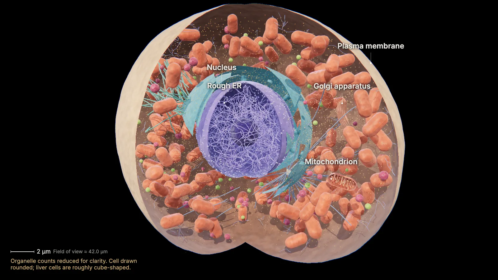
The parts of an animal cell
- Plasma membrane
- Cytoplasm
- Nucleus
- Nuclear pores & nucleolus
- Ribosomes
- Rough endoplasmic reticulum
- Smooth endoplasmic reticulum
- Golgi apparatus
- Lysosomes & peroxisomes
- Mitochondria
- Cytoskeleton & centrosome
| Part | What it does | Size |
|---|---|---|
| Plasma membrane | Separates the cell from its surroundings and controls what goes in and out | Bilayer thickness: ≈ 4 nm |
| Cytoplasm | Gel-like cytosol in which the organelles are suspended; many reactions, including protein synthesis, happen here | Water in cytoplasm: 70–80 % Cytosol (liver cell): 54 % of volume |
| Nucleus | Houses the cell's DNA | Diameter (liver cell): ≈ 8 µm Share of cell volume: ≈ 6 % |
| Nuclear pores & nucleolus | Pores control traffic between nucleus and cytoplasm; the nucleolus assembles ribosomal subunits | Nuclear pore: ≈ 120 nm Nucleoli: 0.5–5 µm |
| Ribosomes | Build proteins | Ribosome diameter: 20–30 nm Large : small subunit: ≈ 3 : 1 |
| Rough endoplasmic reticulum | Studded with ribosomes; new proteins enter it and are folded and modified | Rough ER (liver cell): ≈ 9 % of volume |
| Smooth endoplasmic reticulum | Makes lipids and steroid hormones, detoxifies, stores calcium ions | ER tubule diameter: 30–100 nm |
| Golgi apparatus | Sorts, tags, packages and distributes proteins and lipids | Transport vesicle: 40–80 nm SER + Golgi (liver cell): ≈ 6 % of volume |
| Lysosomes & peroxisomes | Lysosomes digest; peroxisomes break down fatty acids and detoxify | Lysosome: 0.1–1.2 µm, pH 4.5–5 Peroxisome: 0.1–1.0 µm |
| Mitochondria | Make ATP by cellular respiration | Size: ≈ 1 × 2 µm Per rat liver cell: ≈ 1,665 Share of volume (liver): ≈ 22 % |
| Cytoskeleton & centrosome | Microtubules, microfilaments and intermediate filaments give shape and hold organelles; the centrosome organizes microtubules | Centriole: ≈ 0.2 × 0.5 µm Microtubule / actin: ≈ 25 nm / 6 nm |
Where marked, values are for a liver cell (rat), as measured in the sources below.
The nuclear envelope, ER, Golgi, vesicles, lysosomes and plasma membrane work together as one system: see the endomembrane system, with the route a protein takes through it.
Plasma membrane
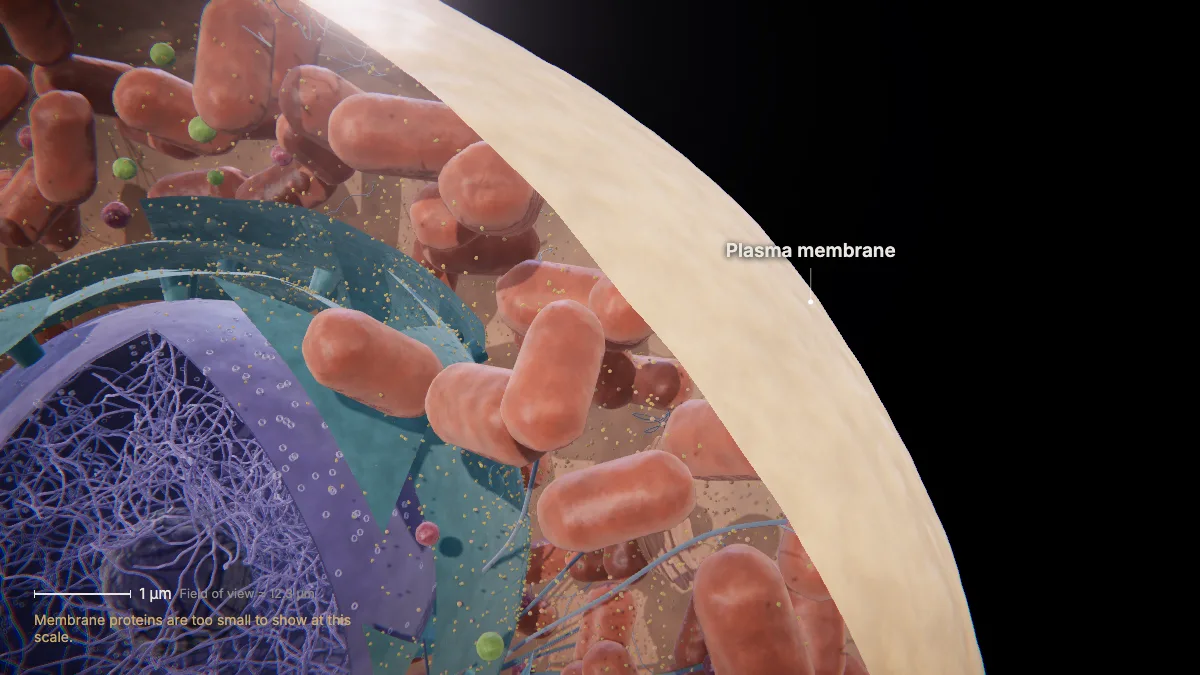
The plasma membrane separates the cell's interior from its surroundings.
It is a double layer of phospholipids with proteins embedded in it.
It controls what passes in and out: organic molecules, ions, water and oxygen. Wastes such as carbon dioxide and ammonia leave through it.
The lipid bilayer is only about 4 nm thick — far too thin to see at this scale.
- Bilayer thickness≈ 4 nm
In the model: Membrane proteins are too small to show at this scale.
Cytoplasm
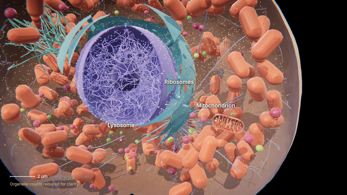
Between the plasma membrane and the nucleus, organelles are suspended in a gel-like cytosol.
The cytoplasm is 70–80 % water. Many metabolic reactions, including protein synthesis, happen here.
In a liver cell, the cytosol alone fills about 54 % of the cell's volume.
- Water in cytoplasm70–80 %
- Cytosol (liver cell)54 % of volume
In the model: Organelle counts reduced for clarity.
Nucleus
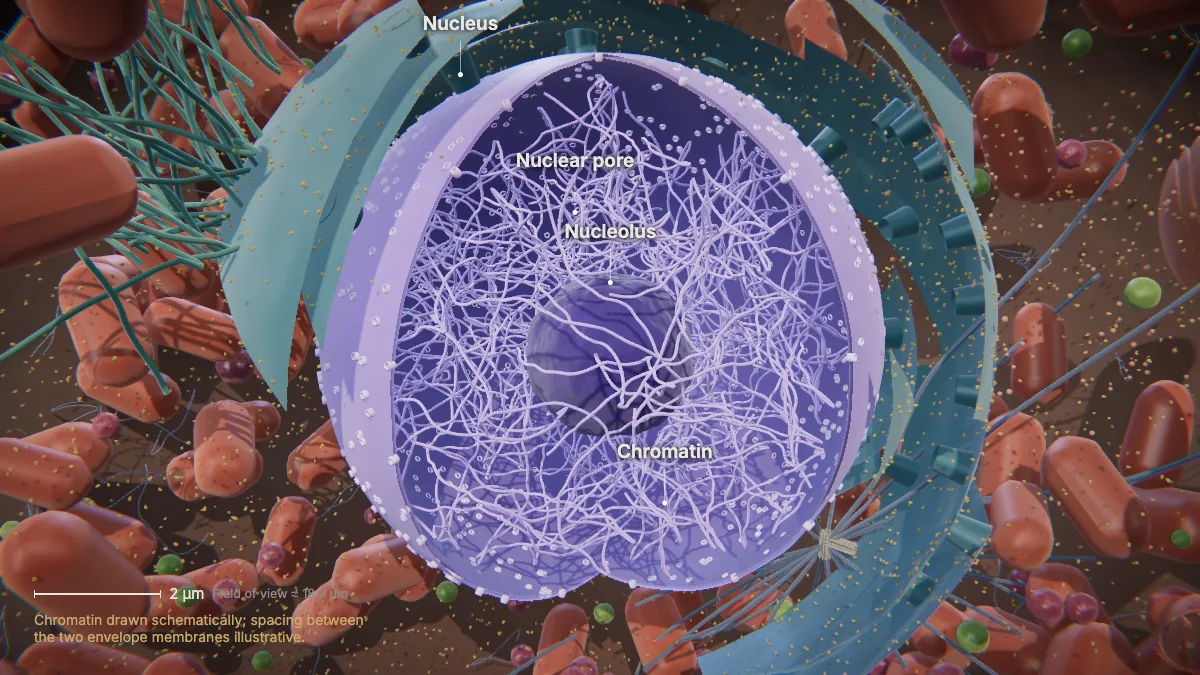
The nucleus houses the cell's DNA and is usually its most prominent organelle.
The nuclear envelope is a double membrane — two phospholipid bilayers — and it is continuous with the endoplasmic reticulum.
Inside, DNA and proteins form chromatin. Human cells have 46 chromosomes.
In a liver cell the nucleus is about 8 µm across and fills roughly 6 % of the cell's volume.
- Diameter (liver cell)≈ 8 µm
- Share of cell volume≈ 6 %
In the model: Chromatin drawn schematically; spacing between the two envelope membranes illustrative.
Nuclear pores & nucleolus
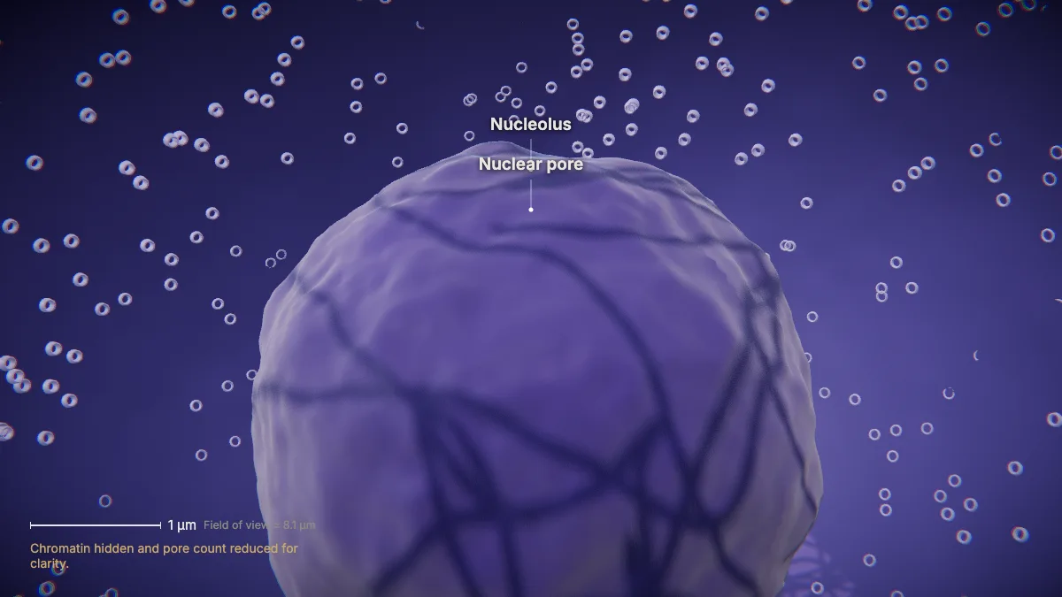
Pores in the envelope control traffic of ions, molecules and RNA between the nucleus and cytoplasm.
Each nuclear pore complex is about 120 nm across.
The dark nucleolus assembles ribosomal RNA with proteins into ribosomal subunits, which leave through the nuclear pores.
- Nuclear pore≈ 120 nm
- Nucleoli0.5–5 µm
In the model: Chromatin hidden and pore count reduced for clarity.
Ribosomes
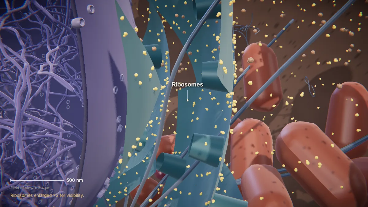
Ribosomes build proteins. Each has a large and a small subunit — the large one about three times bigger.
Some ribosomes float free in the cytoplasm; others are attached to the endoplasmic reticulum or the outer nuclear membrane.
A ribosome is only 20–30 nm wide.
- Ribosome diameter20–30 nm
- Large : small subunit≈ 3 : 1
In the model: Ribosomes enlarged ×2 for visibility.
Rough endoplasmic reticulum
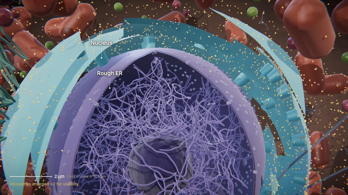
Rough ER is studded with ribosomes. New proteins enter its lumen, where they are folded and modified.
The rough ER also makes phospholipids for cell membranes.
In a liver cell, rough ER cisternae take up about 9 % of the cell's volume.
- Rough ER (liver cell)≈ 9 % of volume
In the model: Ribosomes enlarged ×2 for visibility.
Smooth endoplasmic reticulum
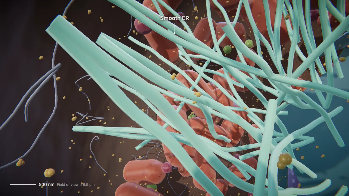
Smooth ER is continuous with the rough ER but has few or no ribosomes.
It makes carbohydrates, lipids and steroid hormones, detoxifies medications and poisons, and stores calcium ions.
ER tubules are only 30–100 nm in diameter.
- ER tubule diameter30–100 nm
Golgi apparatus
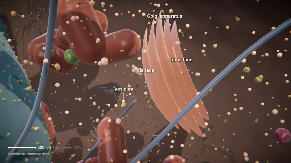
The Golgi sorts, tags, packages and distributes proteins and lipids.
Transport vesicles from the ER fuse with its cis face — the side closer to the ER.
Finished products leave the trans face in secretory vesicles. Some fuse with the plasma membrane and release their contents outside the cell.
- Transport vesicle40–80 nm
- SER + Golgi (liver cell)≈ 6 % of volume
In the model: Number of cisternae simplified.
Lysosomes & peroxisomes
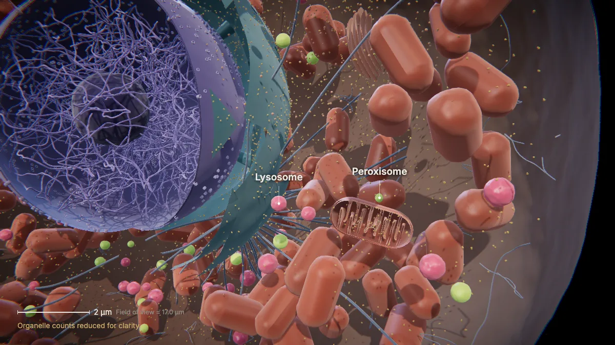
Lysosomes are the cell's digestive compartments; peroxisomes break down fatty acids and detoxify poisons.
Lysosome enzymes break down proteins, polysaccharides, lipids, nucleic acids and even worn-out organelles. Their interior is acidic, about pH 4.5–5.
Peroxisomes are small, round and enclosed by a single membrane. Their enzymes safely break down hydrogen peroxide.
- Lysosome0.1–1.2 µm, pH 4.5–5
- Peroxisome0.1–1.0 µm
In the model: Organelle counts reduced for clarity.
Mitochondria
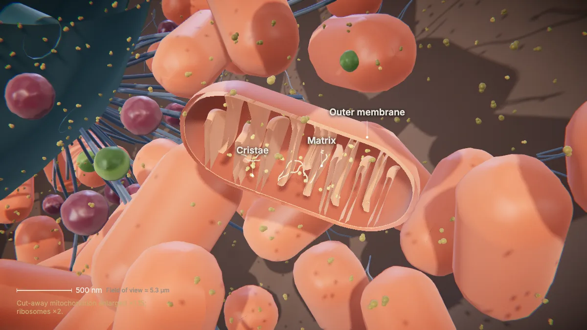
Mitochondria make ATP through cellular respiration, using oxygen and releasing carbon dioxide.
They have a double membrane. The inner membrane folds inward into cristae, which increase its surface area for chemical reactions.
The space inside the inner membrane is the matrix. It holds the mitochondrion's own DNA and ribosomes.
They are about 1 µm wide and 2 µm long. A rat liver cell holds about 1,665 of them; in liver cells they fill about 22 % of the volume.
- Size≈ 1 × 2 µm
- Per rat liver cell≈ 1,665
- Share of volume (liver)≈ 22 %
In the model: Cut-away mitochondrion enlarged ×1.15; ribosomes ×2.
Cytoskeleton & centrosome
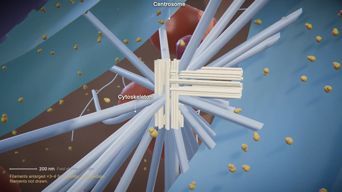
The centrosome near the nucleus organizes microtubules. It holds two centrioles at right angles.
Each centriole is a cylinder of nine triplets of microtubules.
Microtubules keep the cell's shape and form the spindle during division; microfilaments line the membrane as a cortex; intermediate filaments hold organelles in place.
- Centriole≈ 0.2 × 0.5 µm
- Microtubule / actin≈ 25 nm / 6 nm
In the model: Filaments enlarged ×3–4 for visibility. Intermediate filaments not drawn.
Making a model of an animal cell?
Keep the parts in proportion. A rat liver cell is roughly a cube about 15 µm on each edge. Scale everything up 10,000 times and 1 µm becomes 1 cm:
| Part | Real size | At ×10,000 |
|---|---|---|
| Whole cell (edge) | ≈ 15 µm | ≈ 15 cm |
| Nucleus (diameter) | ≈ 8 µm | ≈ 8 cm |
| Mitochondrion | ≈ 1 × 2 µm | ≈ 1 × 2 cm |
| Lysosome | 0.1–1.2 µm | 1–12 mm |
| Ribosome | 20–30 nm | 0.2–0.3 mm |
Ribosomes are far smaller than anything else you will build — at this scale they would be grains of sand. A rat liver cell holds about 1,665 mitochondria, so a model can only show some of them.
For teachers
CytoTour is built for teaching biology in class. Chapter 1, this tour of the cell, is free for everyone.
- Project it and go step by step, with labels and an English voice-over you can turn on or off.
- Checkpoints pause the lesson for a question: predict before a step, or check after it.
- Record a lesson video right from the page, or export an MP4.
- Simplifications are stated on screen, and the scale bar is measured from the actual view.
The Teacher plan adds the next chapters — transcription, translation, and protein folding and secretion — plus 4K video exports without a watermark: $29 a year during early access (regular price $69).
What the model simplifies
- Organelle counts are reduced so you can see inside; a toggle in the app shows the true density of mitochondria.
- Ribosomes are drawn 2× and cytoskeleton filaments 3–4× their real size so they stay visible, and the opened mitochondrion is enlarged 1.15×. Each of these views says so.
- The number of Golgi cisternae is illustrative.
- The cell is drawn rounded; liver cells are roughly cube-shaped. Its volume is kept close to a real liver cell's.
- Colors are illustrative — organelles are not these colors in real life.
Sources
- OpenStax, Biology 2e, §4.3 Eukaryotic Cells — openstax.org
- OpenStax, Biology 2e, §4.4 The Endomembrane System and Proteins — openstax.org
- OpenStax, Microbiology, §3.4 Unique Characteristics of Eukaryotic Cells — openstax.org
- Alberts et al., Molecular Biology of the Cell, 5th ed., Table 12-1 (organelle volumes in a liver cell) — via BioNumbers
- Milo & Phillips, Cell Biology by the Numbers — book.bionumbers.org
- Harvard BioNumbers database (sizes of individual organelles) — bionumbers.hms.harvard.edu