 CYTOTOUR
CytoTour › Transcription and translation
Transcription and translation: protein synthesis in 3D
A gene is first transcribed into RNA in the nucleus; the processed mRNA then carries its information to ribosomes in the cytoplasm, where it is translated into a chain of amino acids. This guide follows one human gene — β-globin (HBB) — through every step, with labeled 3D views built from real molecular structures.
From gene to protein
- DNA (gene)
- pre-mRNA
- mRNA (cap, tail, introns removed)
- Nuclear pore
- Ribosome
- Polypeptide
Transcription vs translation
| Transcription | Translation | |
|---|---|---|
| Where (in eukaryotes) | In the nucleus (RNA polymerase II is located there) | On ribosomes in the cytoplasm and on the surface of the rough ER |
| What is read | One DNA strand — the template strand — read 3'→5' | mRNA, read 5'→3' in triplets called codons |
| What is made | RNA, built 5'→3' (pre-mRNA, then processed into mRNA) | A polypeptide, built from the N terminus to the C terminus |
| Main machine | RNA polymerase II, with transcription factors | The ribosome (rRNA + proteins), with tRNAs carrying amino acids |
| Pairing rule | Each RNA base pairs with a template base: template A → RNA U, T → A, G → C, C → G | tRNA anticodons pair with mRNA codons |
| Start signal | The promoter (here with a TATA-like box) upstream of +1 | The start codon AUG (methionine) |
| End signal | Pol II runs on 1,000–2,000 nt past the gene; the RNA is then cut between an AAUAAA signal and a GU-rich sequence | A stop codon: UAA, UAG or UGA |
| Speed (measured) | ≈ 18–72 nucleotides per second | ≈ 5 amino acids per second (human) |
Speeds are measured values from the BioNumbers database; transcription speed varies by gene and cell.
Transcription, step by step
Inside the nucleus, DNA wraps around histone proteins as nucleosomes. Here we zoom in on one gene: human β-globin (HBB).
These views are Chapter 2 of CytoTour, part of the Teacher plan.
The gene: DNA in the nucleus
1Into the nucleus
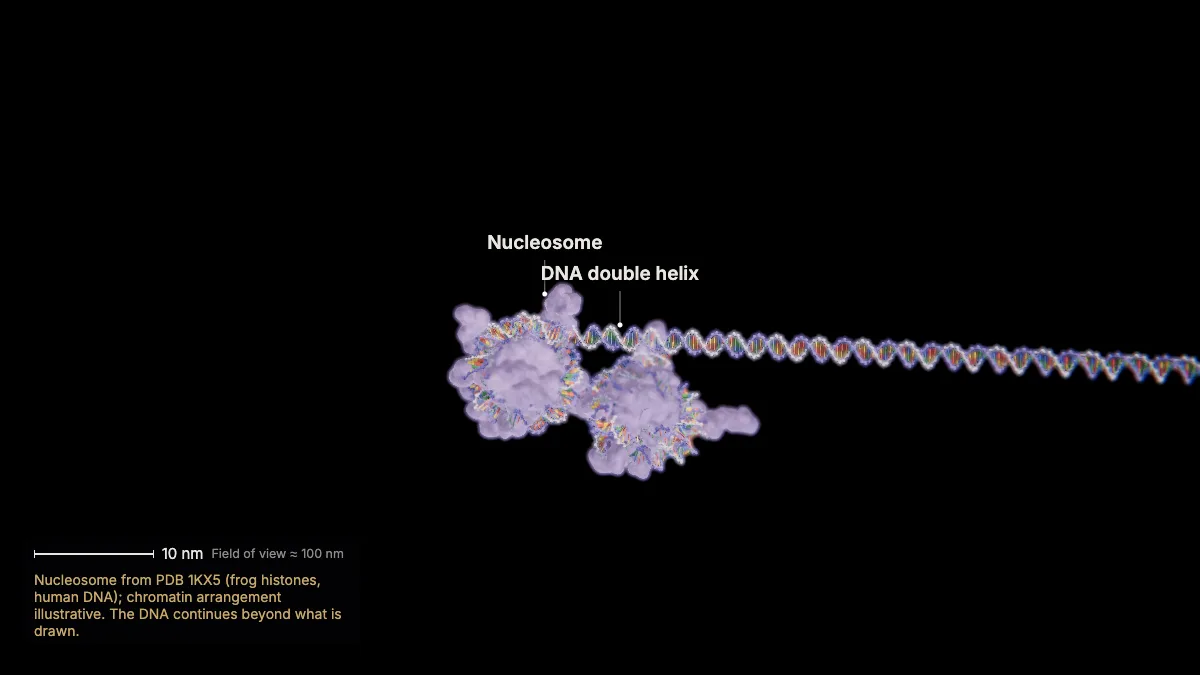
Inside the nucleus, DNA wraps around histone proteins as nucleosomes. Here we zoom in on one gene: human β-globin (HBB).
Each nucleosome wraps about 146 base pairs of DNA around a core of histones, and nucleosomes are regularly spaced along the DNA.
A nucleosome is a disc roughly 10 nm across. The molecules in this chapter are built at true scale from their experimental structures.
- DNA per nucleosome≈ 146 bp
- Nucleosome≈ 10 nm
In the model: Nucleosome from PDB 1KX5 (frog histones, human DNA); chromatin arrangement illustrative. The DNA continues beyond what is drawn.
2The double helix
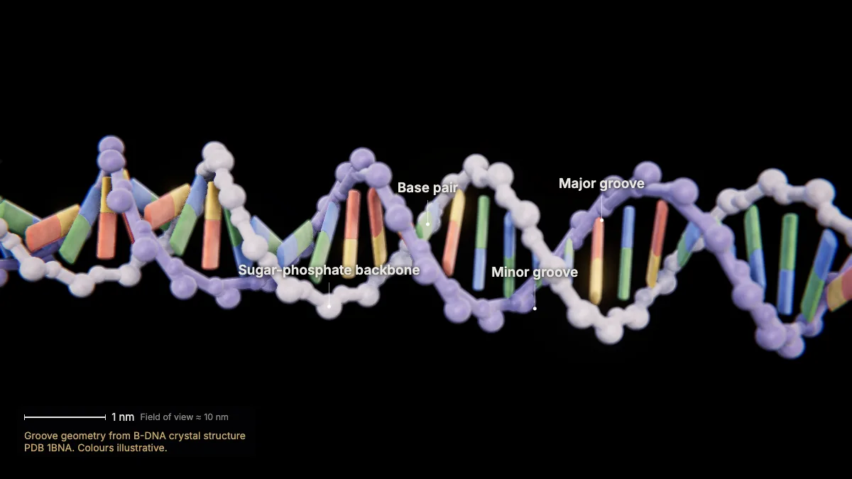
Two antiparallel strands twist into a right-handed helix, 2 nm wide, with 10 base pairs per turn.
Sugar and phosphate form the backbone on the outside; the bases stack inside like the rungs of a ladder.
A pairs with T through two hydrogen bonds; G pairs with C through three. Each pair joins a double-ring purine to a single-ring pyrimidine.
The twist creates major and minor grooves — binding sites for DNA-binding proteins.
- Diameter2 nm
- Rise per base pair0.34 nm
- Per turn10 bp · 3.4 nm
In the model: Groove geometry from B-DNA crystal structure PDB 1BNA. Colours illustrative.
Initiation: finding the start
3The β-globin promoter
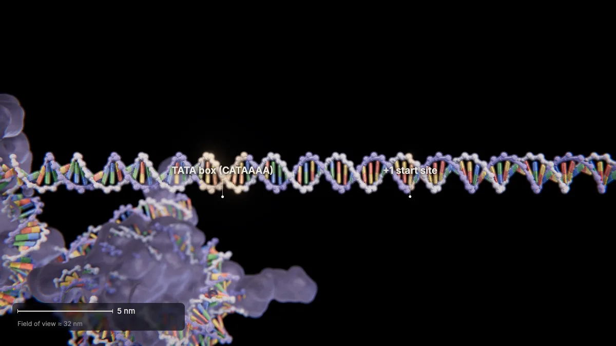
Upstream of the gene, the promoter carries a TATA box — here the TATA-like sequence CATAAAA at −32 to −26.
TATA boxes (consensus TATAAA on the coding strand) sit about 25–35 bases upstream of the +1 start site.
The HBB gene has 3 exons and 2 introns, spanning 1,608 nucleotides from the +1 start site to the end of exon 3.
- TATA window−25 to −35
- HBB exons / introns3 / 2
4TFIID finds the TATA box
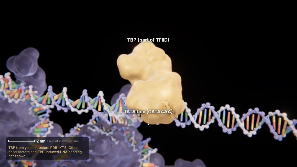
Basal transcription factors bind the promoter first. The core factor, TFIID, contains the TATA-binding protein (TBP).
RNA polymerase and transcription factors bind promoter or enhancer sequences to start transcription.
Together the basal factors build a preinitiation complex on the DNA.
In the model: TBP from yeast structure PDB 1YTB. Other basal factors and TBP-induced DNA bending not shown.
5RNA polymerase II arrives
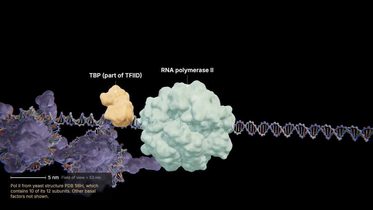
RNA polymerase II binds to form the transcription initiation complex. In the nucleus, it makes all protein-coding pre-mRNAs.
Pol II is a large molecular machine — yeast Pol II is built from a dozen different proteins.
Measured from its crystal structure, it is about 15 × 14 × 12 nm — some 7 times wider than the DNA it reads.
- Pol II size (PDB 1I6H)≈ 15 × 14 × 12 nm
In the model: Pol II from yeast structure PDB 1I6H, which contains 10 of its 12 subunits. Other basal factors not shown.
Elongation: copying the template strand
6Opening the bubble
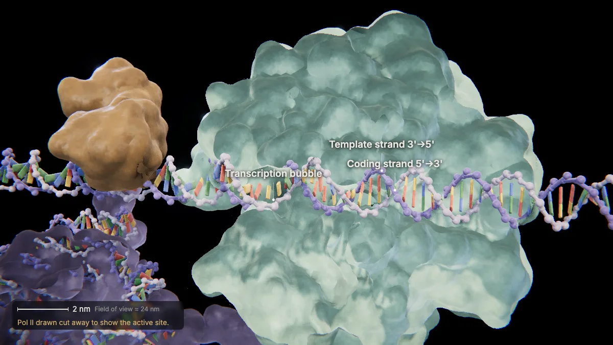
At the start site the DNA is unwound, opening a transcription bubble of about 15 base pairs.
Only one of the two strands — the template strand — will be read.
Pol II is cut away here so you can see inside the active site.
- Bubble≈ 15 bp
In the model: Pol II drawn cut away to show the active site.
7Elongation
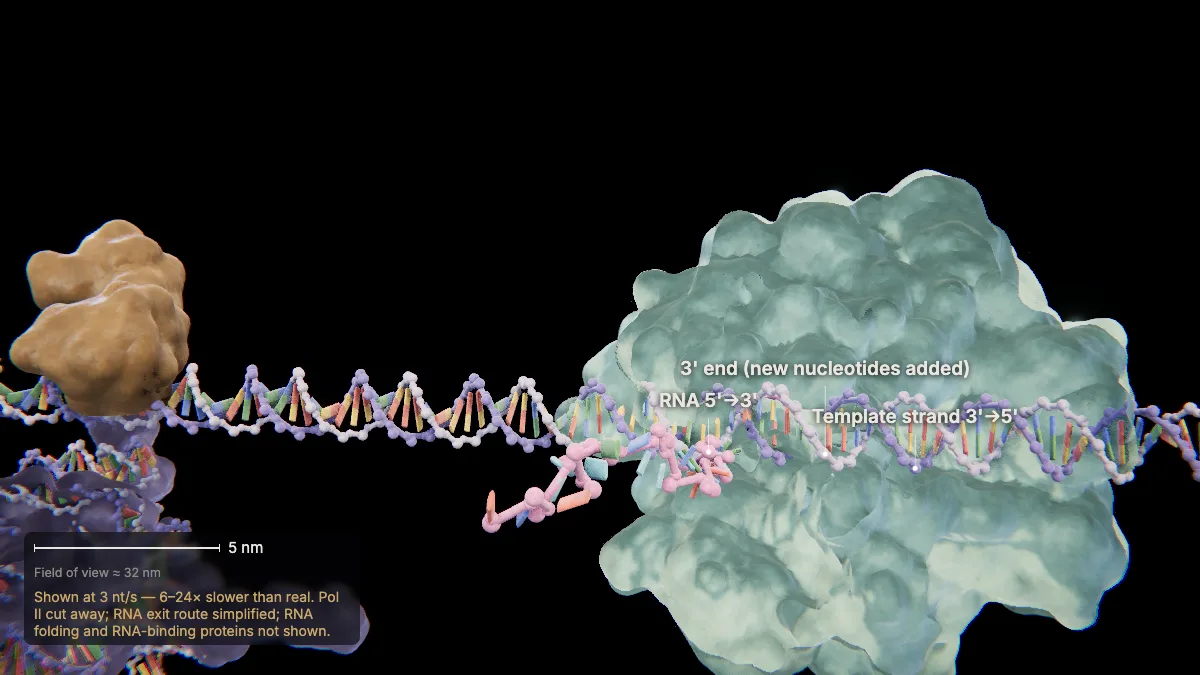
Pol II reads the template strand 3'→5' and builds RNA 5'→3', adding each new nucleotide to the 3' end.
Each new RNA base pairs with the template base: template A with RNA U, T with A, G with C, and C with G.
Ahead of the enzyme the DNA is unwound; behind it, it rewinds. Only the newest 9–10 RNA nucleotides stay paired with the template.
Real Pol II runs at roughly 18–72 nucleotides per second — slowed down here.
In the model: Shown at 3 nt/s — 6–24× slower than real. Pol II cut away; RNA exit route simplified; RNA folding and RNA-binding proteins not shown.
8Template vs coding strand
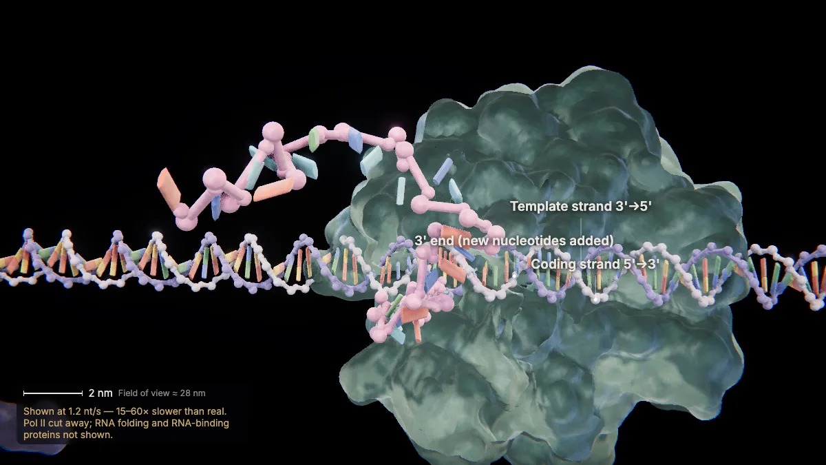
The RNA is complementary to the template strand — so it reads the same as the coding strand, with U in place of T.
Compare the rows in the sequence panel: coding strand and RNA match letter for letter, except every T becomes U.
That is why the coding strand is also called the nontemplate strand.
In the model: Shown at 1.2 nt/s — 15–60× slower than real. Pol II cut away; RNA folding and RNA-binding proteins not shown.
RNA processing: cap, tail and splicing
9Capping the 5' end
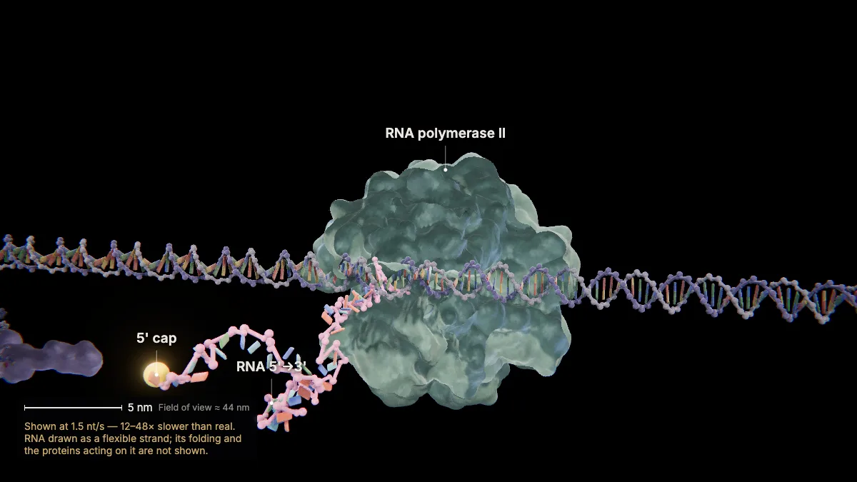
While the RNA is still being made, a 7-methylguanosine cap is added to its 5' end.
The cap protects the new RNA from being degraded.
Later, protein-synthesis factors recognize the cap to help ribosomes start translation.
In the model: Shown at 1.5 nt/s — 12–48× slower than real. RNA drawn as a flexible strand; its folding and the proteins acting on it are not shown.
10To the end of the gene
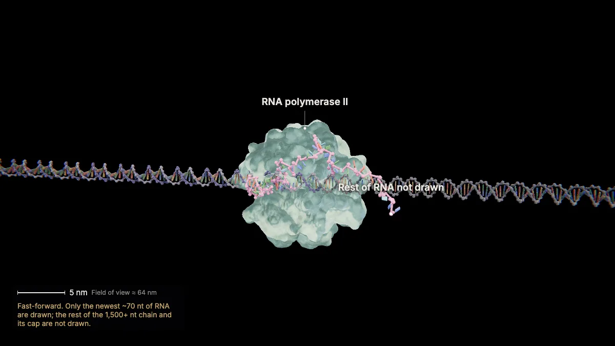
Fast-forward: Pol II copies all 1,608 nucleotides of HBB — exons and introns alike — and runs on 1,000–2,000 nucleotides before stopping.
The whole primary transcript, introns included, is called pre-mRNA.
The extra read-through RNA beyond the gene is cut off during processing.
In the model: Fast-forward. Only the newest ~70 nt of RNA are drawn; the rest of the 1,500+ nt chain and its cap are not drawn.
11Cleavage and the poly-A tail
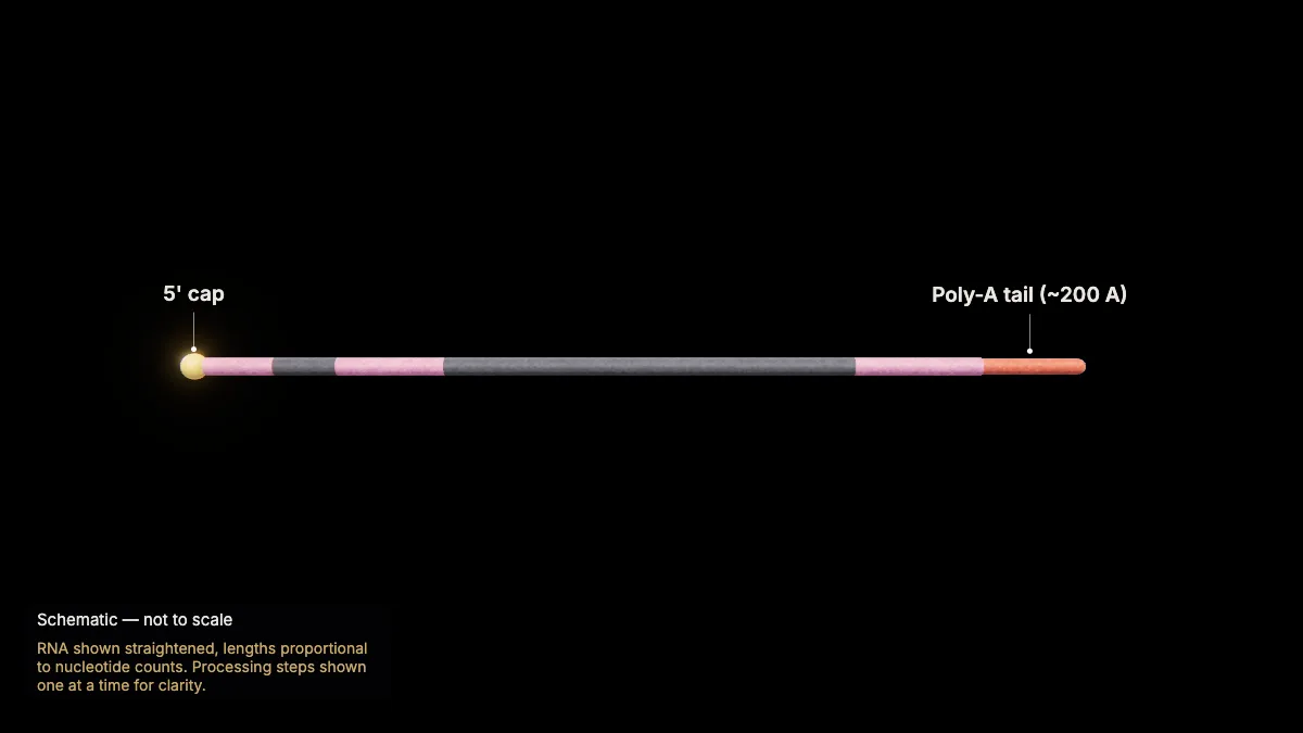
The pre-mRNA is cut between an AAUAAA signal and a GU-rich sequence; poly-A polymerase then adds about 200 adenines.
HBB's AAUAAA signal sits near the end of exon 3.
The poly-A tail makes the mRNA more stable and binds a protein needed to export it from the nucleus.
In the model: RNA shown straightened, lengths proportional to nucleotide counts. Processing steps shown one at a time for clarity.
12Splicing out the introns
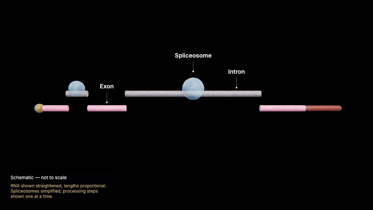
Spliceosomes — complexes of proteins and small nuclear RNAs — cut out both introns and join the three exons.
Exons carry the protein-coding sequence; introns are removed while the RNA is still in the nucleus.
Each intron begins with GU and ends with AG. Every intron must be removed completely and precisely.
Joining exons in different combinations can produce different mature mRNAs — alternative splicing.
- Introns removed130 + 850 nt
- Mature mRNA628 nt + cap + tail
In the model: RNA shown straightened, lengths proportional. Spliceosomes simplified; processing steps shown one at a time.
Export to the cytoplasm
13Out through a nuclear pore
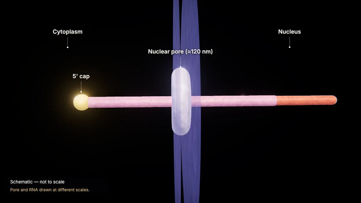
The finished mRNA leaves the nucleus through a nuclear pore, heading for ribosomes in the cytoplasm.
Nuclear pores, about 120 nm across, regulate the movement of ions, molecules and RNA between the nucleus and the cytoplasm.
Next chapter: a ribosome reads this mRNA and builds β-globin protein.
In the model: Pore and RNA drawn at different scales.
Translation, step by step
These views are Chapter 3 of CytoTour, part of the Teacher plan. The genetic code is also on our free codon chart.
The message, the code and the machines
14A message in the cytoplasm
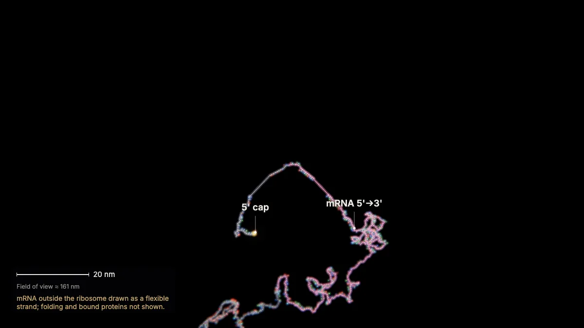
The finished β-globin mRNA has left the nucleus. In the cytoplasm, ribosomes translate it into a chain of amino acids.
mRNA carries information from DNA in the nucleus to ribosomes in the cytoplasm. Translation happens on ribosomes in the cytoplasm and on the surface of the rough ER.
This mRNA is 628 nucleotides plus a cap and a poly-A tail; its coding sequence begins at nucleotide 51.
In the model: mRNA outside the ribosome drawn as a flexible strand; folding and bound proteins not shown.
15The genetic code
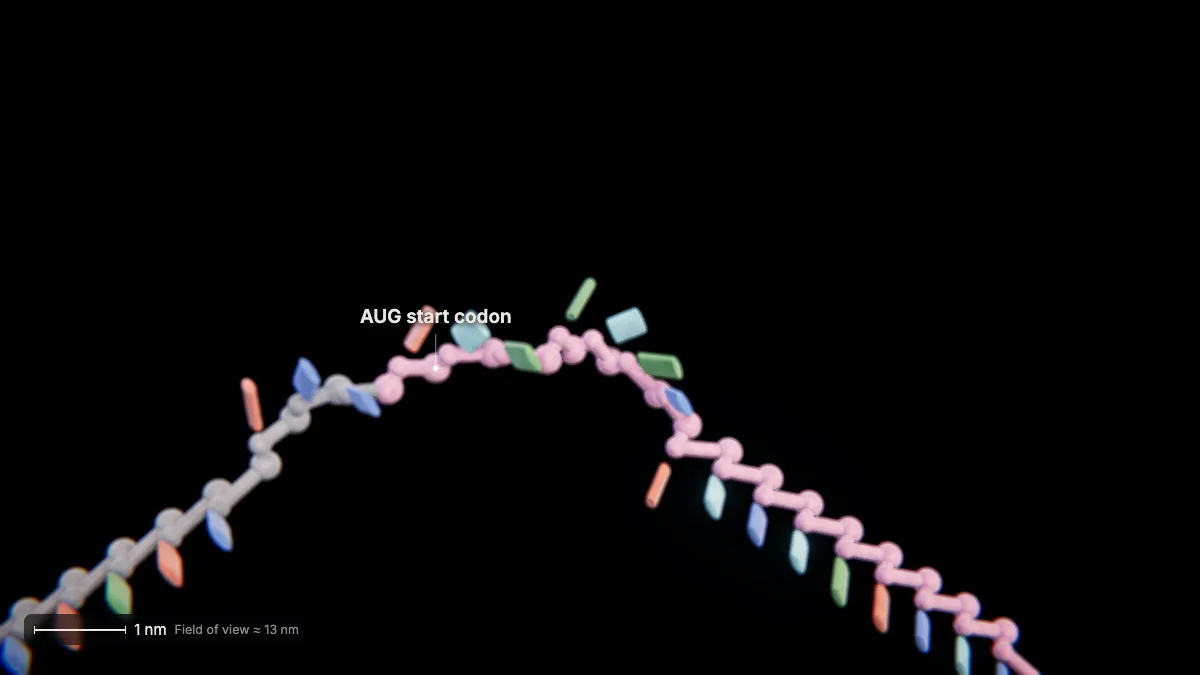
The mRNA is read in triplets called codons. AUG — methionine — is the start codon and sets the reading frame.
There are 64 codons: 61 specify amino acids and 3 (UAA, UAG, UGA) are stop codons. Most amino acids have more than one codon.
Proteins are built from 20 common amino acids, and the code is colinear: nucleotides 1–3 give amino acid 1, and so on.
Nearly all living things use the same genetic code — evidence of common ancestry.
16The ribosome
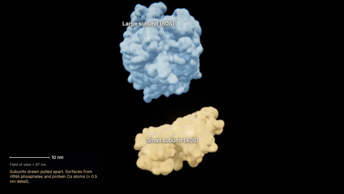
A ribosome is built from ribosomal RNA and many proteins, in two subunits: small (40S) and large (60S), 80S together.
The small subunit binds the mRNA; the large subunit binds tRNAs.
This human ribosome, from its cryo-EM structure (PDB 9T6C), measures about 30 × 27 × 22 nm.
- Human 80S (PDB 9T6C)≈ 30 × 27 × 22 nm
In the model: Subunits drawn pulled apart. Surfaces from rRNA phosphates and protein Cα atoms (≈ 0.5 nm detail).
17Transfer RNA
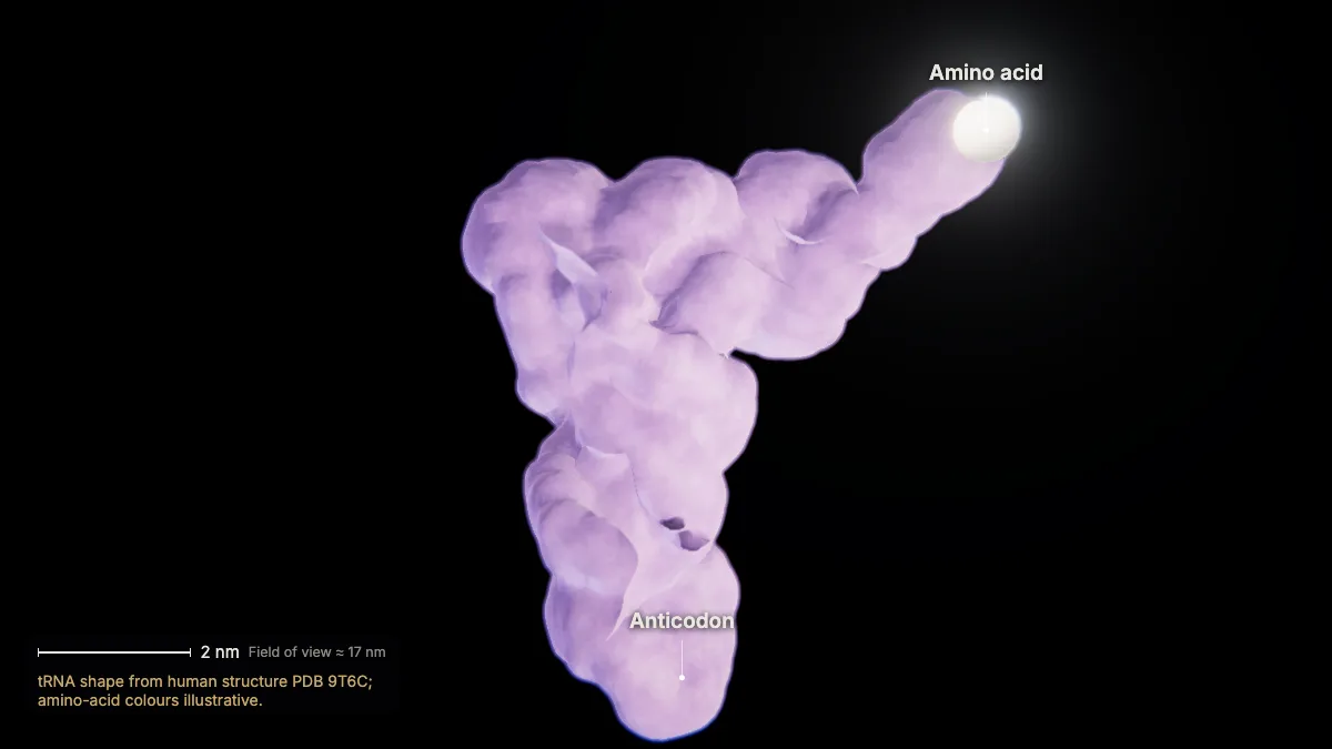
Each tRNA is a short RNA folded into an L shape: an anticodon at one end, its amino acid at the other.
The anticodon base-pairs with an mRNA codon for that tRNA's amino acid.
Enzymes called aminoacyl-tRNA synthetases charge each tRNA with its correct amino acid, using energy from ATP.
Cells have 40–60 kinds of tRNA, depending on the species.
- tRNA length70–90 nt
In the model: tRNA shape from human structure PDB 9T6C; amino-acid colours illustrative.
Initiation
18Initiation
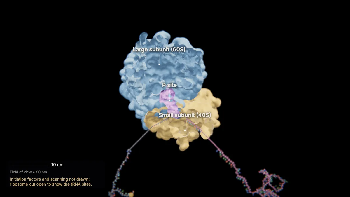
The small subunit, with initiation factors, recognizes the 5' cap and moves along the mRNA to the start codon. Then the large subunit joins.
The initiator tRNA carrying methionine sits in the P site, paired with AUG.
HBB's start codon sits in a classic context: …ACACC AUG G… (A at −3, G at +4).
In the model: Initiation factors and scanning not drawn; ribosome cut open to show the tRNA sites.
Elongation: one codon at a time
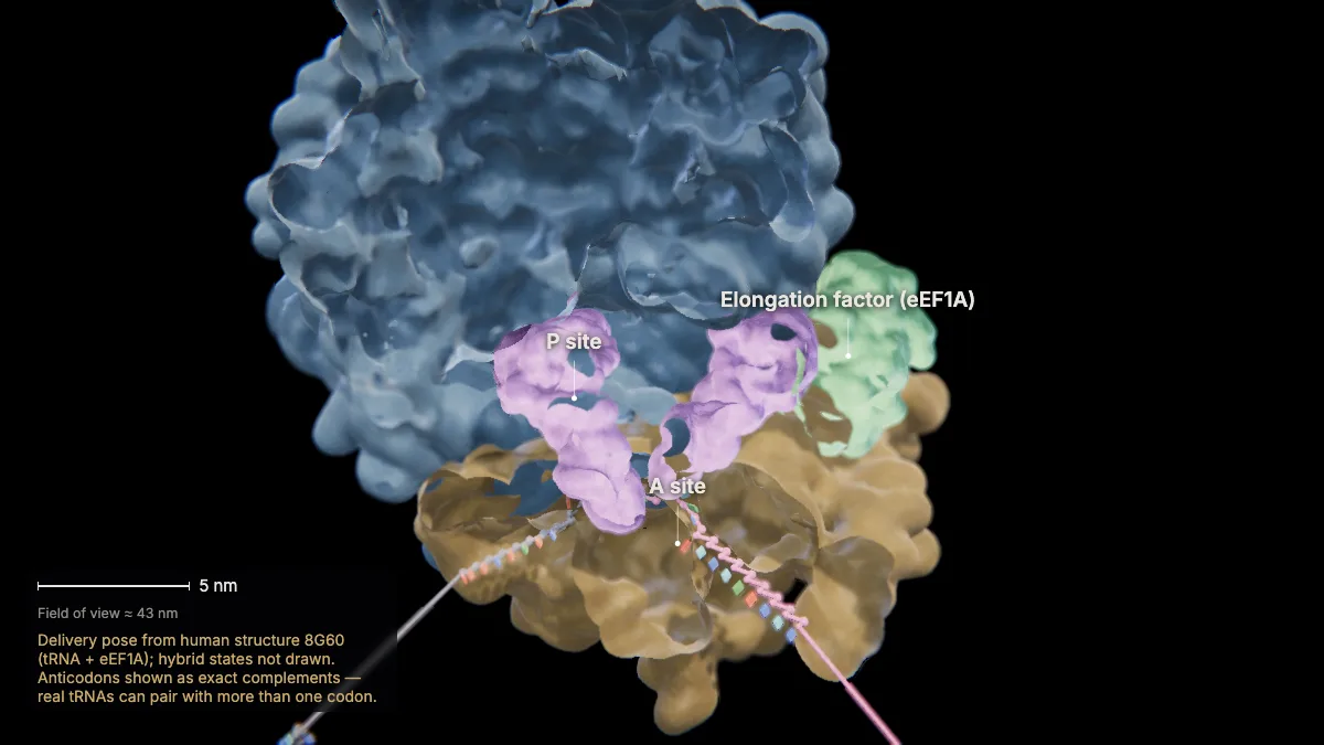
19A new tRNA arrives
An elongation factor delivers the next charged tRNA into the A site. Codon 2 is GUG, so it brings valine.
The tRNA's anticodon must pair with the A-site codon — that is how the correct amino acid is brought to each codon.
The A site receives incoming tRNAs; the P site holds the tRNA with the growing chain; the E site releases empty tRNAs.
In the model: Delivery pose from human structure 8G60 (tRNA + eEF1A); hybrid states not drawn. Anticodons shown as exact complements — real tRNAs can pair with more than one codon.
20Forming a peptide bond
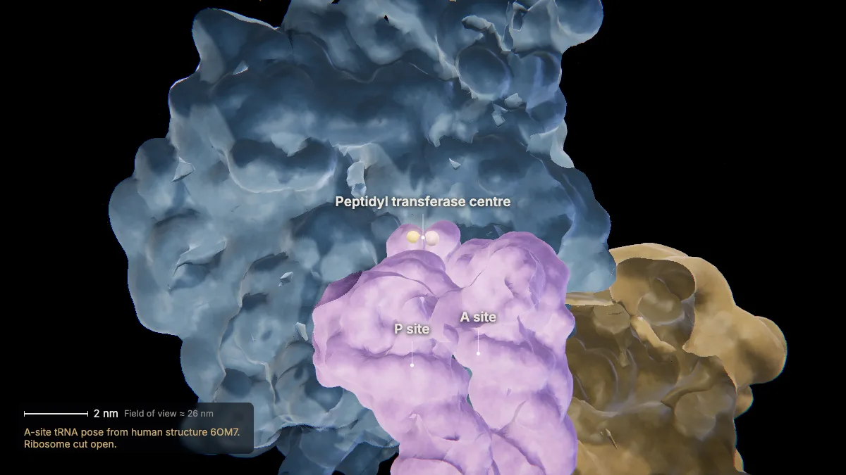
The ribosome joins the growing chain to the new amino acid. The catalyst is RNA — the ribosome is a ribozyme.
Peptidyl transferase, an RNA-based enzyme in the large subunit, forms the peptide bond.
The energy comes from the high-energy bond that linked the amino acid to its tRNA.
In the model: A-site tRNA pose from human structure 6OM7. Ribosome cut open.
21Translocation
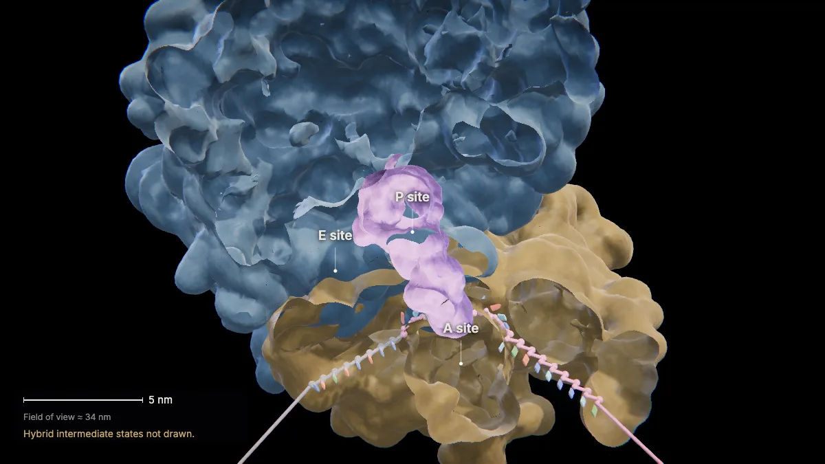
The ribosome moves three nucleotides toward the mRNA's 3' end. The tRNAs shift from A to P and from P to E.
The empty tRNA leaves from the E site to be recharged.
Elongation factors that hydrolyze GTP power each step.
In the model: Hybrid intermediate states not drawn.
22Elongation continues
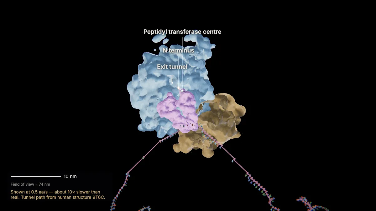
The cycle repeats codon by codon. The chain grows from its N terminus to its C terminus and threads out through a tunnel in the large subunit.
Human ribosomes add about 5 amino acids per second.
The exit tunnel is roughly 8–10 nm long.
In the model: Shown at 0.5 aa/s — about 10× slower than real. Tunnel path from human structure 9T6C.
23One letter, one amino acid
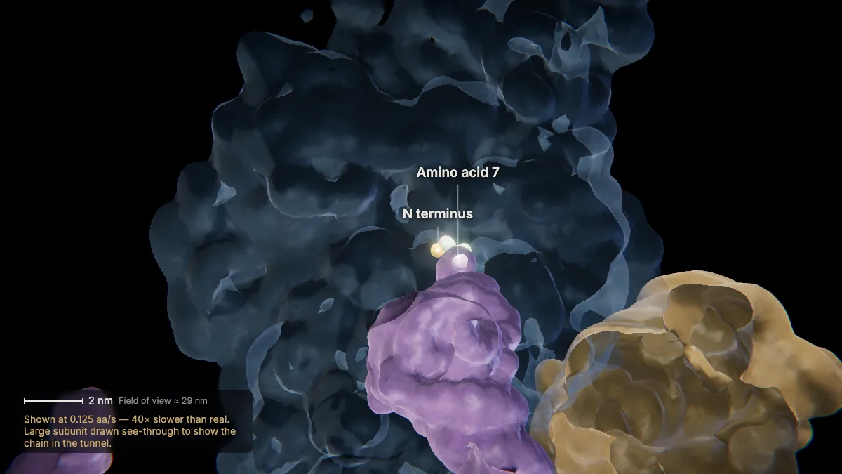
Amino acid 7 is glutamate, from codon GAG. In sickle-cell hemoglobin a single base change makes it GUG — valine.
A point mutation substitutes one nucleotide for another. Use the HbS switch in Conditions to see it.
Counting from the mature protein, whose first methionine is removed, this is position 6 (β6).
Carrying one HbS copy confers resistance to malaria — a genetic change that natural selection can act on.
In the model: Shown at 0.125 aa/s — 40× slower than real. Large subunit drawn see-through to show the chain in the tunnel.
Termination
24To the stop codon
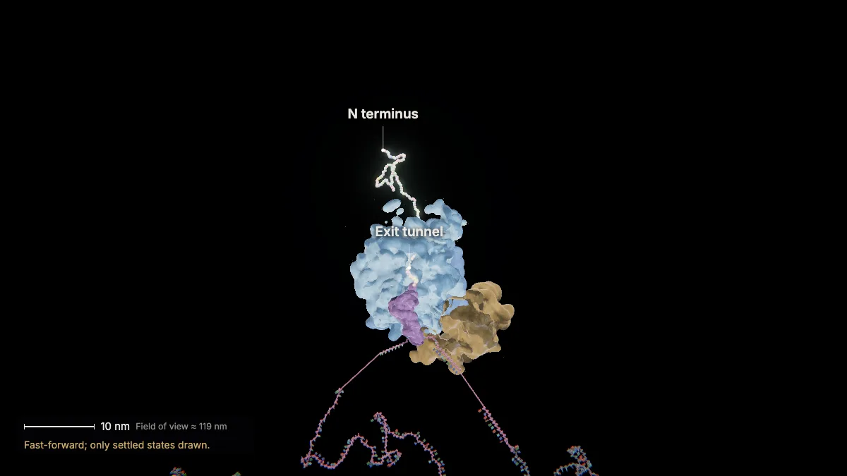
Fast-forward through all 147 codons until the stop codon UAA reaches the A site.
The three stop codons (UAA, UAG, UGA) code for no amino acid. HBB ends with UAA.
In the model: Fast-forward; only settled states drawn.
25Termination
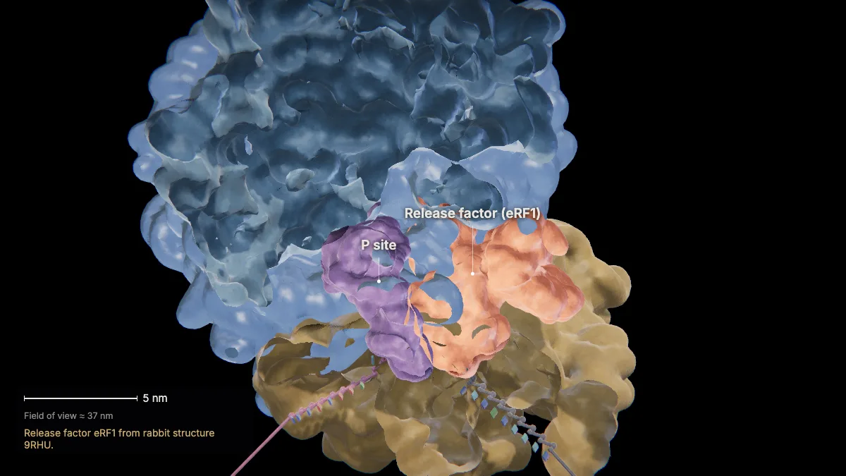
A protein release factor — shaped like a tRNA — binds the stop codon. Water is added to the chain's end and the protein is released.
Translation continues along the mRNA until a stop codon is reached, then ends with the release of the new protein.
In the model: Release factor eRF1 from rabbit structure 9RHU.
26Subunits separate
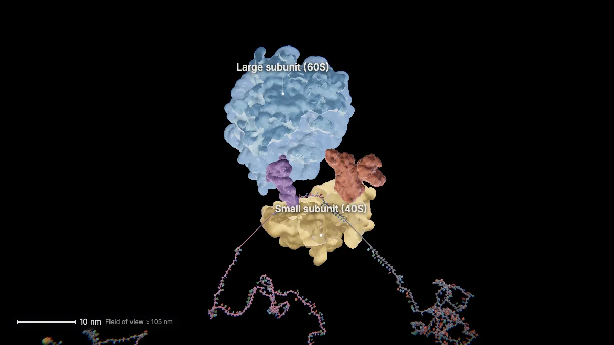
Factors using energy from GTP separate the large and small subunits from the mRNA.
After translation
27Many ribosomes at once
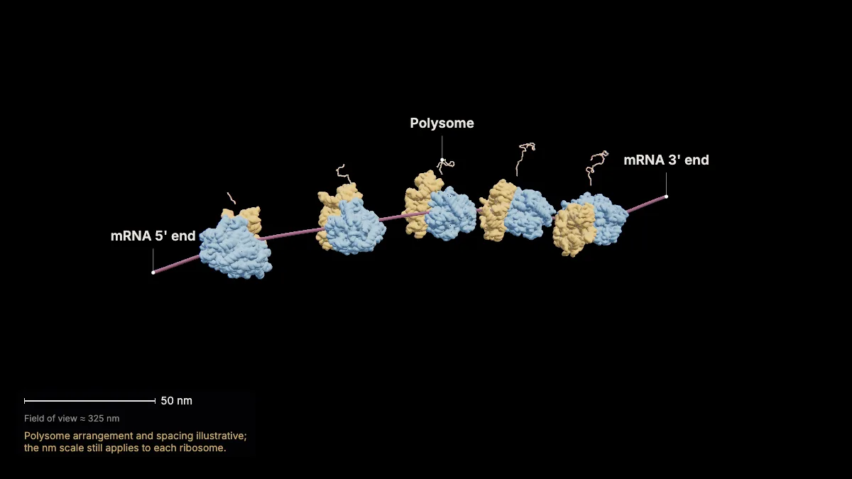
Each mRNA is read by many ribosomes at the same time — a polysome. Chains are longer on ribosomes nearer the 3' end.
In the model: Polysome arrangement and spacing illustrative; the nm scale still applies to each ribosome.
28A new chain
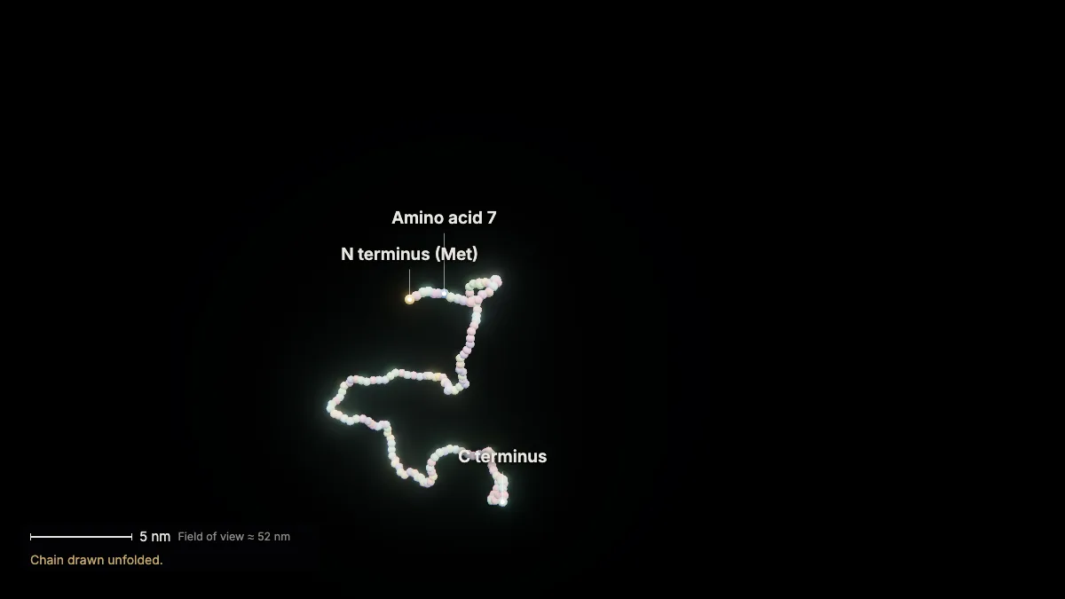
The finished chain has 147 amino acids. Its first methionine is removed, leaving the 146-residue β-globin chain.
During and after translation, amino acids can be modified, and some proteins need helper chaperones to fold.
Next chapter: how new chains fold, and how proteins made on the rough ER are sent through the cell.
In the model: Chain drawn unfolded.
For teachers
These chapters follow the AP Biology Course and Exam Description topics on transcription and RNA processing (Topic 6.3), translation (Topic 6.4) and mutations (Topic 6.7, with sickle-cell anemia as the example).
- Project the 3D model and go step by step, with labels and an English voice-over you can turn on or off.
- Checkpoints pause the lesson for a question: predict before a step, or check after it.
- Record a lesson video right from the page, or export an MP4.
- Simplifications are stated on screen, and the scale bar is measured from the actual view.
Chapter 1, the tour of the cell, is free for everyone. The Teacher plan adds the next chapters — transcription, translation, and protein folding and secretion — plus 4K video exports without a watermark: $29 a year during early access (regular price $69).
What the model simplifies
- Structures come from several species: RNA polymerase II and TBP are yeast proteins, the nucleosome has frog histones, and the release factor is from rabbit; the gene, ribosome and tRNAs are human. Each view says which.
- Only TBP of the transcription factors is drawn, and the DNA bending it causes is not shown.
- RNA polymerase II and the ribosome are drawn cut open to show their active sites. One tRNA shape is reused for all tRNAs, and intermediate (hybrid) states are not drawn.
- Most steps run far slower than real time, with fast-forwards in between; the on-screen readout gives the factor.
- RNA processing is shown on a straightened RNA map, with lengths proportional to nucleotide counts, one step at a time.
- Colors are illustrative.
Sources
- OpenStax, Biology 2e, §14.2 DNA Structure and Sequencing — openstax.org
- OpenStax, Biology 2e, §15.1–15.5 Genes and Proteins (the genetic code, transcription, RNA processing, ribosomes and protein synthesis) — openstax.org
- OpenStax, Microbiology, §11.3 RNA Transcription — openstax.org
- College Board, AP Biology Course and Exam Description, Topics 6.3, 6.4, 6.6, 6.7 — apcentral.collegeboard.org
- Ensembl, HBB-201 transcript (ENST00000335295), GRCh38 — ensembl.org; UniProt P68871 (β-globin) — uniprot.org
- NCBI, The Genetic Codes, Table 1 (standard code) — ncbi.nlm.nih.gov
- Lodish et al., Molecular Cell Biology, 8th ed., and Ashkenazy-Titelman et al., Nature Communications 13:5881 (2022) — mRNA leaves the nucleus 5' end first — doi.org
- RCSB PDB-101, Molecules of the Month: RNA Polymerase, Transfer RNA, The Ribosome — pdb101.rcsb.org
- Protein Data Bank structures 1KX5, 1BNA, 1YTB, 1I6H, 9T6C, 6OM7, 8G60, 9RHU — rcsb.org
- Harvard BioNumbers database (rates, sizes) — bionumbers.hms.harvard.edu; Milo & Phillips, Cell Biology by the Numbers — book.bionumbers.org