 CYTOTOUR
CytoTour › Endomembrane system
The endomembrane system: an interactive 3D guide
The endomembrane system is a group of membranes and organelles in eukaryotic cells that works together to modify, package and transport lipids and proteins. This guide shows each part in a labeled 3D model, then follows one protein — insulin — all the way through it.
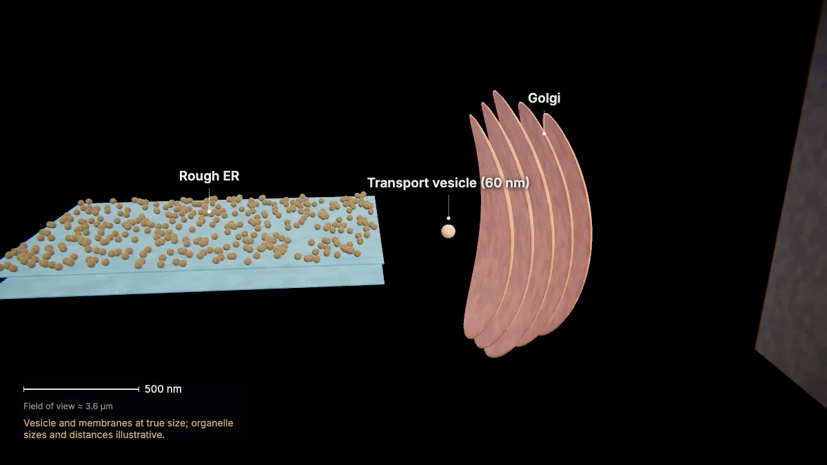
What is part of the endomembrane system?
It includes the nuclear envelope, the endoplasmic reticulum (rough and smooth), the Golgi apparatus, vesicles, lysosomes and the plasma membrane.
| Part | Role |
|---|---|
| Nuclear envelope | Double membrane around the nucleus; continuous with the ER |
| Rough endoplasmic reticulum (rough ER) | Ribosomes on its surface; new proteins enter it and are folded and modified |
| Smooth endoplasmic reticulum (smooth ER) | Makes lipids and steroid hormones, detoxifies, stores calcium ions |
| Golgi apparatus | Sorts, tags, packages and distributes proteins and lipids |
| Vesicles | Carry proteins from the ER to the Golgi, and from the Golgi onward |
| Lysosomes | Digest and recycle; destroy pathogens |
| Plasma membrane | Bounds the cell; secretory vesicles fuse with it to release their contents |
Not part of it
Mitochondria and chloroplasts have membranes of their own, but they are not part of the endomembrane system.
The parts, one by one
Nuclear envelope
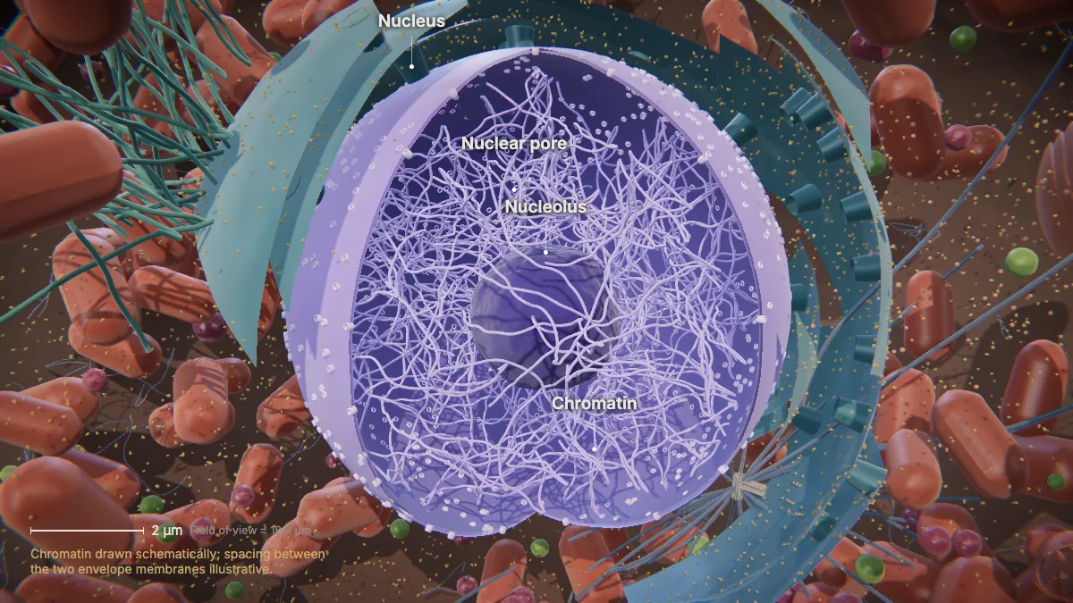
The nuclear envelope is a double membrane — two phospholipid bilayers — and it is continuous with the endoplasmic reticulum.
Rough endoplasmic reticulum (rough ER)
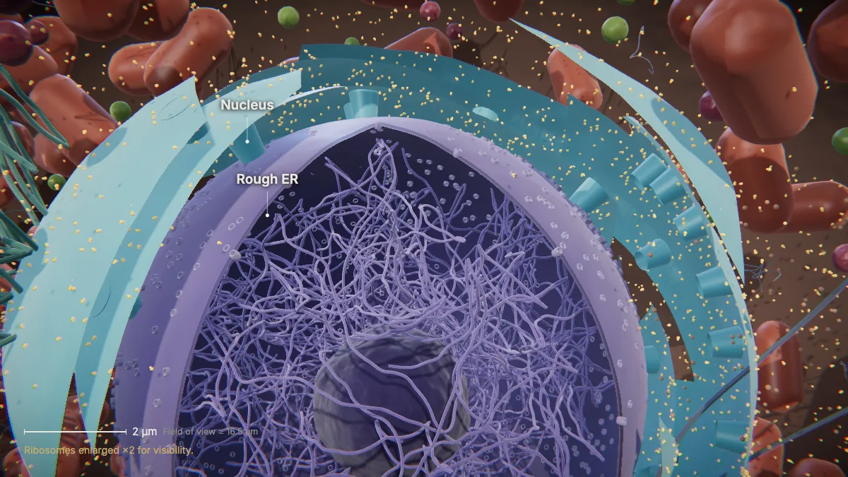
Rough ER is studded with ribosomes. New proteins enter its lumen, where they are folded and modified.
The rough ER also makes phospholipids for cell membranes.
Smooth endoplasmic reticulum (smooth ER)
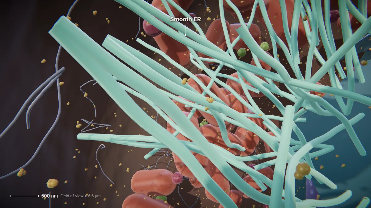
Smooth ER is continuous with the rough ER but has few or no ribosomes.
It makes carbohydrates, lipids and steroid hormones, detoxifies medications and poisons, and stores calcium ions.
Golgi apparatus
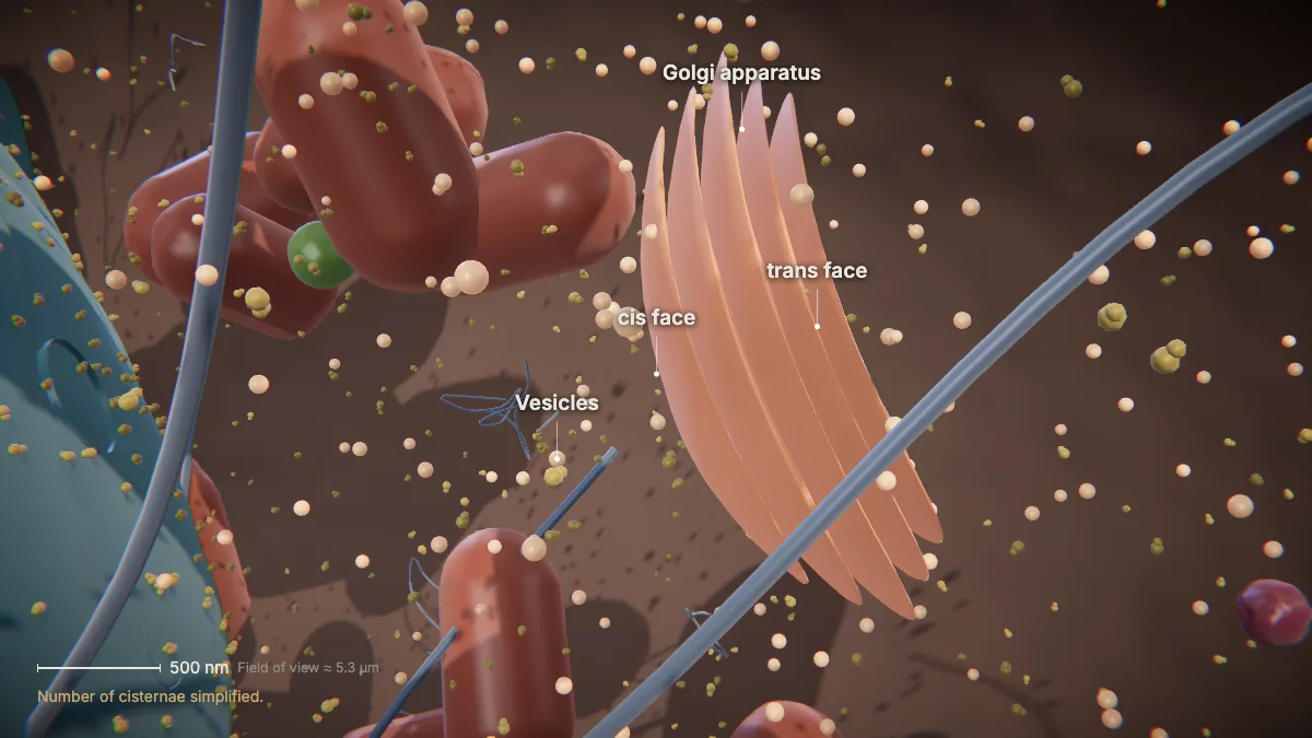
The Golgi sorts, tags, packages and distributes proteins and lipids.
Transport vesicles from the ER fuse with its cis face — the side closer to the ER.
Finished products leave the trans face in secretory vesicles. Some fuse with the plasma membrane and release their contents outside the cell.
Vesicles
Transport vesicles bud from the ER and carry proteins to the cis face of the Golgi; secretory vesicles bud from the Golgi's trans face.
A transport vesicle is only about 40–80 nm across.
Lysosomes
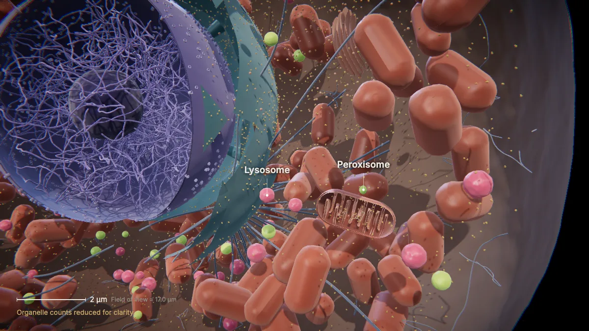
Lysosomes are the cell's digestive and recycling compartments. Their enzymes also destroy pathogens that enter the cell.
Lysosome enzymes break down proteins, polysaccharides, lipids, nucleic acids and even worn-out organelles. Their interior is acidic, about pH 4.5–5.
Plasma membrane
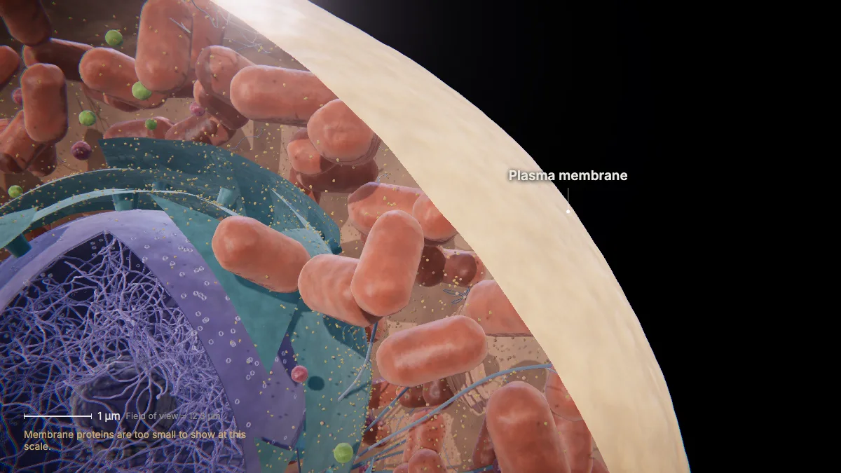
The plasma membrane separates the cell's interior from its surroundings.
It is a double layer of phospholipids with proteins embedded in it.
Follow a protein through it: insulin
Insulin is made by the β-cells of the pancreas and secreted into the blood, where it lowers blood glucose.
Insulin is made for export, so it takes the secretory route through the endomembrane system:
- Rough ER
- Transport vesicle
- Golgi (cis → trans)
- Secretory granule
- Plasma membrane
- Outside the cell
These steps are Chapter 4 of CytoTour, part of the Teacher plan.
1Made as preproinsulin
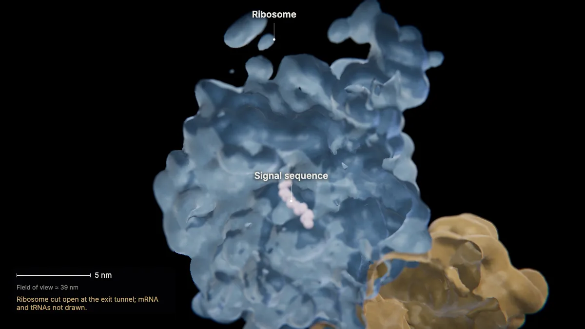
Insulin starts as one 110-amino-acid chain: a signal peptide, then the B chain, a C-peptide, and the A chain.
Preproinsulin is made by ribosomes on the rough ER. The N terminus — the signal peptide — emerges from the ribosome first.
A signal sequence is a short stretch at the amino end that sends a protein to a particular compartment. Secreted proteins carry one.
In the model: Ribosome cut open at the exit tunnel; mRNA and tRNAs not drawn.
2The signal is recognized
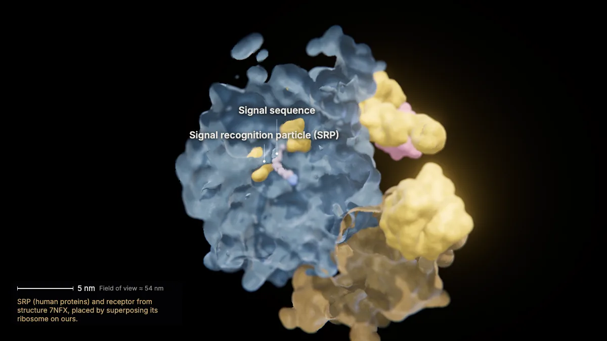
As soon as the signal sequence emerges from the ribosome, the signal recognition particle (SRP) binds it.
Signal-recognition proteins such as SRP act as conductors for proteins headed to the ER.
In the model: SRP (human proteins) and receptor from structure 7NFX, placed by superposing its ribosome on ours.
3Docking at the ER
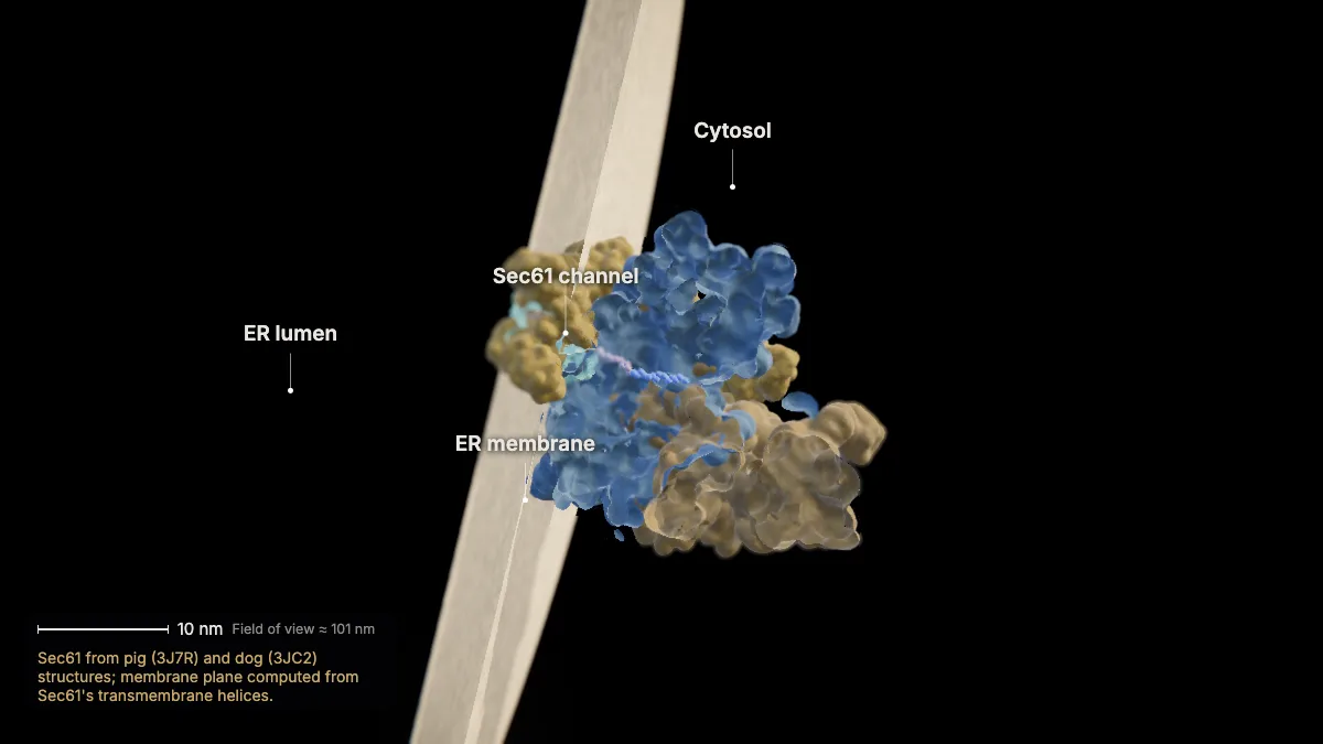
The ribosome docks on a protein channel in the ER membrane — Sec61. The signal sequence opens the channel.
Sec61's main subunit crosses the membrane 10 times. The lipid bilayer itself is only about 4 nm thick.
Rough ER is rough because of these membrane-bound ribosomes.
In the model: Sec61 from pig (3J7R) and dog (3JC2) structures; membrane plane computed from Sec61's transmembrane helices.
4Into the ER lumen
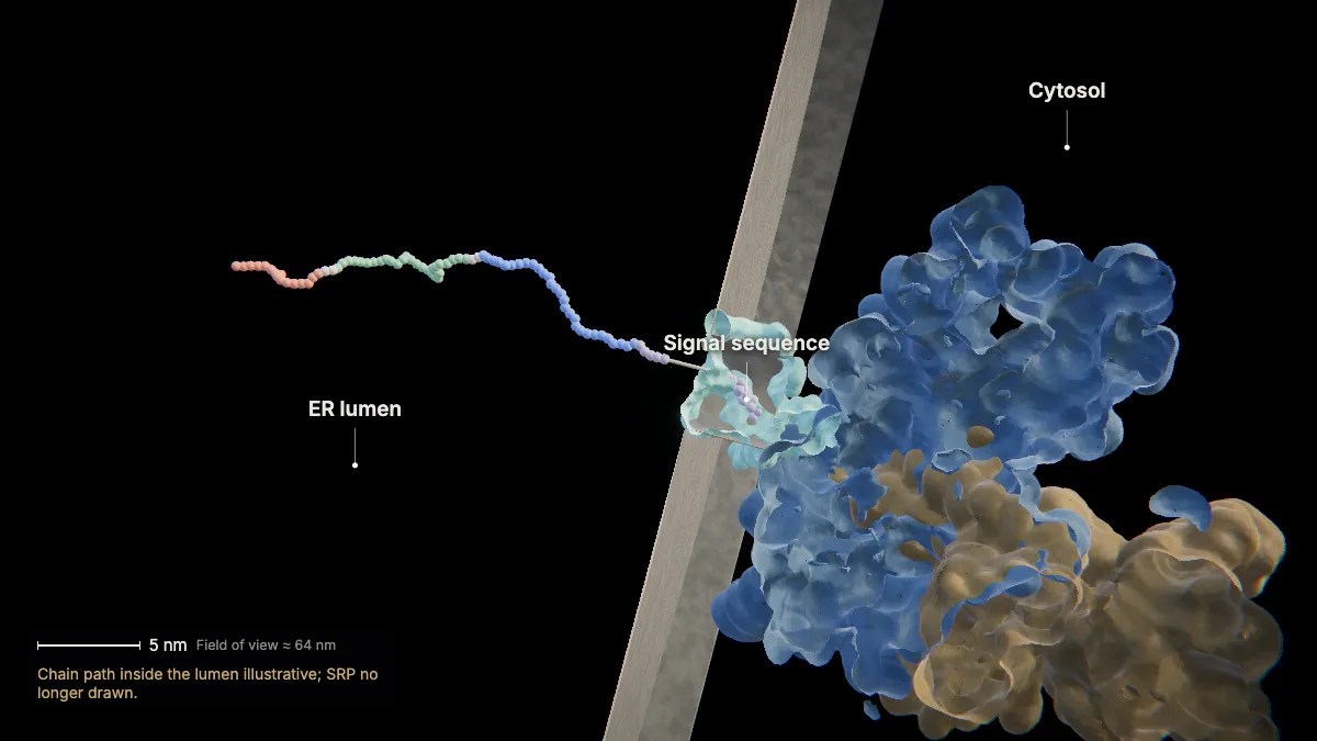
Translation continues, and the growing chain passes straight from the ribosome, through the channel, into the ER lumen.
In the channel structure, the signal sequence sits in the side of the opened channel, and the rest of the chain enters the lumen.
In the model: Chain path inside the lumen illustrative; SRP no longer drawn.
5The signal peptide is removed
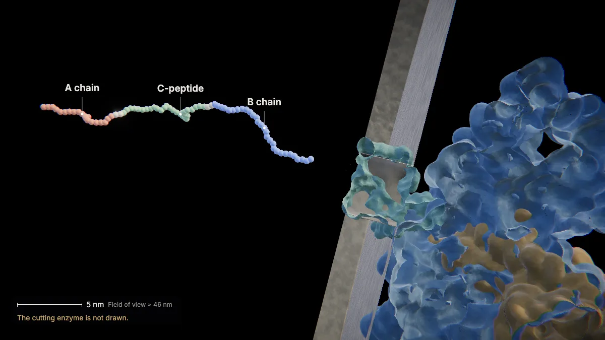
Once inside the ER, the signal peptide is clipped off. What remains is proinsulin: B chain, C-peptide, A chain.
Preproinsulin (110 amino acids) becomes proinsulin (86 amino acids).
In the model: The cutting enzyme is not drawn.
6Folding and disulfide bonds
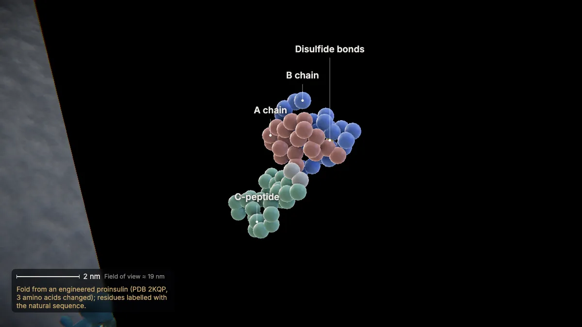
Proinsulin folds in the ER. Three disulfide bonds lock its shape: two link the future A and B chains, one lies within the A chain.
A tertiary shape comes from hydrogen bonds, hydrophobic and ionic interactions, and disulfide bridges.
Many proteins fold on their own; some need helper proteins called chaperones to avoid clumping.
In the model: Fold from an engineered proinsulin (PDB 2KQP, 3 amino acids changed); residues labelled with the natural sequence.
7From the ER to the Golgi
Proinsulin leaves the ER in a transport vesicle, which fuses with the cis face of the Golgi.
Together, the ER, Golgi, vesicles and plasma membrane — the endomembrane system — modify, package and transport proteins.
In the model: Vesicle and membranes at true size; organelle sizes and distances illustrative.
8Packed into granules
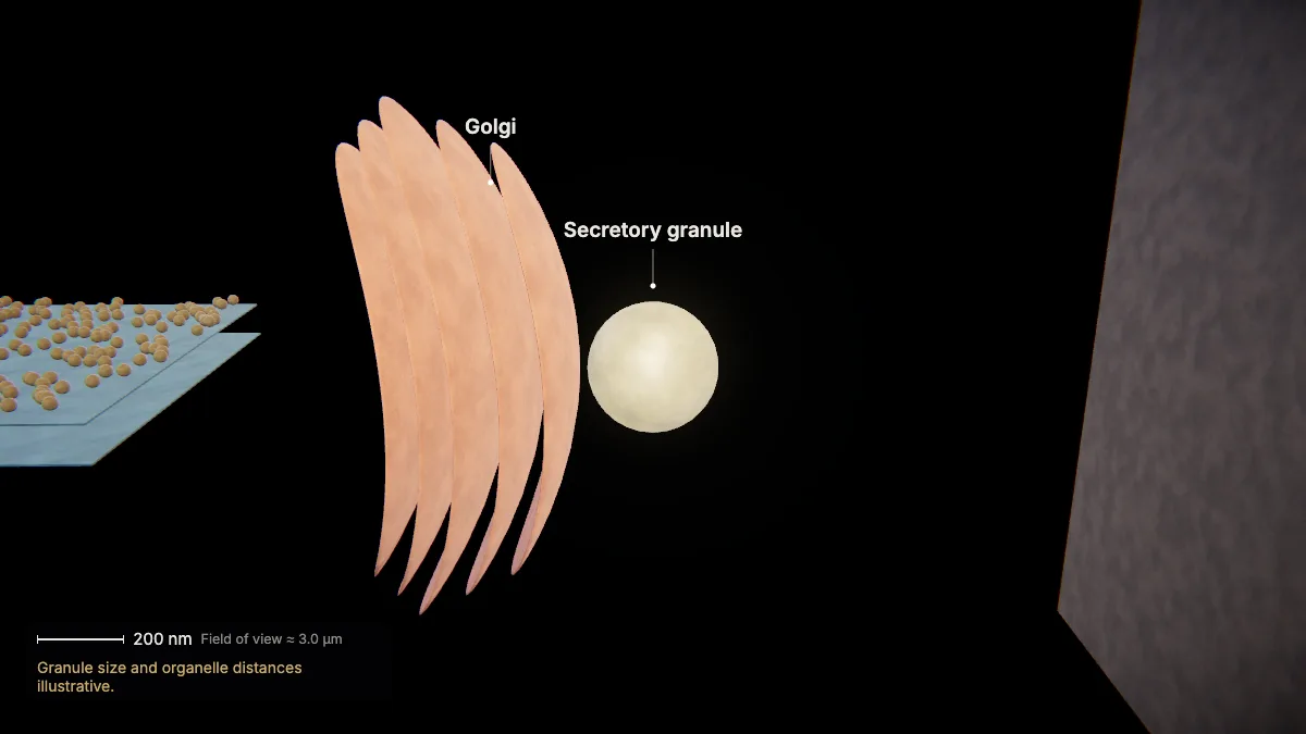
Passing through the Golgi's flattened sacs, proinsulin is packaged at the trans side into immature secretory granules.
The Golgi helps fold and chemically modify new proteins and packages them for trafficking.
In the model: Granule size and organelle distances illustrative.
9Granules mature
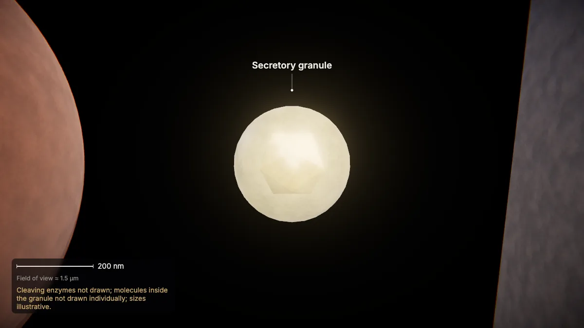
Inside the granule, proinsulin is cut into insulin and C-peptide, and insulin crystallizes with zinc into a dense core.
So the cell builds one longer chain, lets it fold, then cuts away the extra piece — leaving two chains.
One granule holds about a million insulin molecules; a β-cell holds about 10,000 granules.
In the model: Cleaving enzymes not drawn; molecules inside the granule not drawn individually; sizes illustrative.
10Exocytosis
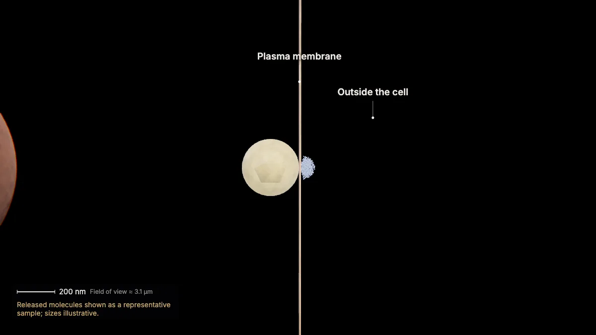
The granule fuses with the plasma membrane and releases insulin outside the cell — exocytosis, which requires energy.
From here insulin enters the blood and lowers blood glucose.
In the model: Released molecules shown as a representative sample; sizes illustrative.
For teachers
This topic is in the AP Biology Course and Exam Description: the endomembrane system, rough ER and Golgi (EK 2.1.A.2–2.1.A.4) and exocytosis (EK 2.5.B.1).
- Project the 3D model and go step by step, with labels and an English voice-over you can turn on or off.
- Checkpoints pause the lesson for a question: predict before a step, or check after it.
- Record a lesson video right from the page, or export an MP4.
- Simplifications are stated on screen, and the scale bar is measured from the actual view.
Chapter 1, the tour of the cell, is free for everyone. The Teacher plan adds the next chapters — transcription, translation, and protein folding and secretion (insulin's route above) — plus 4K video exports without a watermark: $29 a year during early access (regular price $69).
What the model simplifies
- In the insulin route, organelle sizes and the distances between organelles are illustrative; vesicles and membranes keep their true size. The secretory granule's size is illustrative too.
- The roughly one million insulin molecules in a granule are not drawn individually, and the enzymes that cut the chain are not drawn.
- The signal recognition particle is shown only while it targets the ribosome to the ER.
- Molecular structures come from several species (human, rabbit, pig and dog proteins); the proinsulin fold is from an engineered variant with 3 amino acids changed. Each view says which.
- In the whole-cell views, ribosomes are drawn 2× their real size, organelle counts are reduced, and colors are illustrative.
Sources
- OpenStax, Biology 2e, §4.4 The Endomembrane System and Proteins — openstax.org
- OpenStax, Biology 2e, §4.3 Eukaryotic Cells — openstax.org
- OpenStax, Biology 2e, §15.5 Ribosomes and Protein Synthesis — openstax.org
- OpenStax, Biology 2e, §5.4 Bulk Transport — openstax.org
- College Board, AP Biology Course and Exam Description — apcentral.collegeboard.org
- UniProt P01308, human insulin — uniprot.org
- Hou, Min & Pessin, Insulin granule biogenesis, trafficking and exocytosis, Vitamins and Hormones 80 (2009) — Europe PMC
- RCSB PDB-101, Molecule of the Month: Insulin — pdb101.rcsb.org
- Protein Data Bank structures 7NFX, 3J7R, 3JC2, 2KQP, 3W7Y, 4F0N — rcsb.org
- Harvard BioNumbers database (vesicle and organelle sizes, granule counts) — bionumbers.hms.harvard.edu