 CYTOTOUR
CytoTour › Mitosis
The stages of mitosis, in 3D
Mitosis passes a complete genome from a parent cell to two genetically identical daughter cells. This guide follows a human cell — all 46 of its chromosomes — through the cell cycle, every stage of mitosis and cytokinesis.
The stages at a glance
| Stage | What happens |
|---|---|
| Interphase (G1, S, G2) | In G1 the cell grows and duplicates organelles; in S it replicates its DNA; in G2 it makes proteins and ATP to prepare for division. |
| Prophase | The chromosomes condense and become visible, the nucleolus disappears, and the nuclear envelope starts to break up. |
| Prometaphase | The nuclear envelope breaks into pieces. At its centromere, each sister chromatid forms a kinetochore that binds spindle microtubules. |
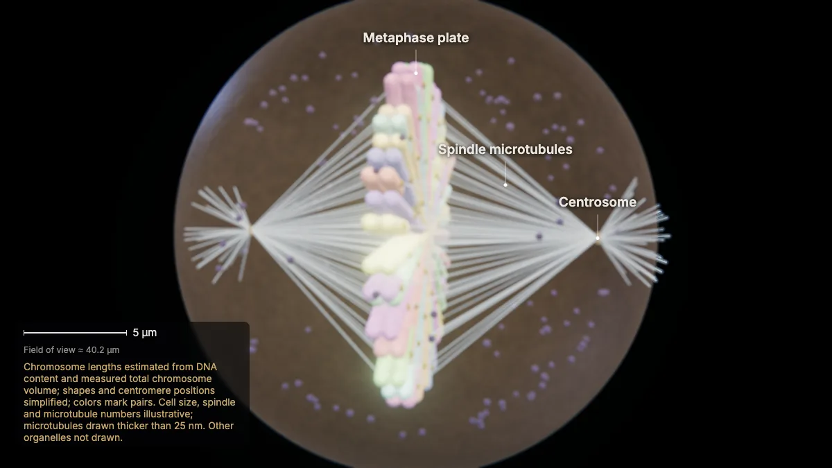
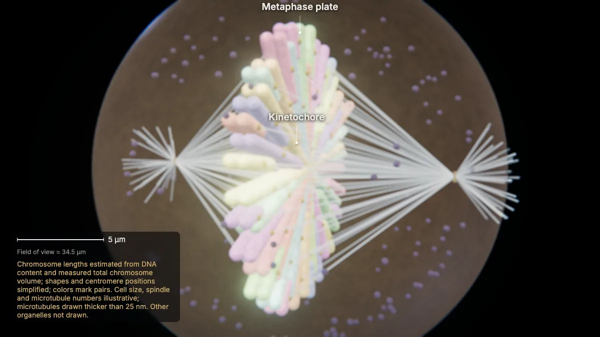
| Metaphase | All the chromosomes line up in one plane at the cell's equator — the metaphase plate. |
| Anaphase | Cohesin breaks down and the sister chromatids separate, pulled to opposite poles. Each chromatid is now its own chromosome. |
| Telophase | The chromosomes reach the poles and decondense; the spindle breaks down, and a nuclear envelope forms around each set. |
| Cytokinesis | A ring of actin filaments just inside the plasma membrane contracts and pinches the cell in two along a cleavage furrow. |
Most of the cell cycle is spent in interphase.
Step by step, in 3D
These views are Chapter 6 of CytoTour, part of the Teacher plan.
The cell cycle
1The cell cycle
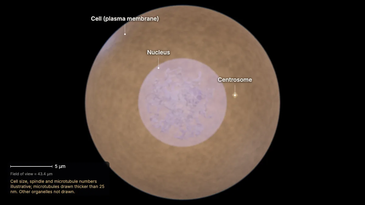
The cell cycle is a tightly regulated series of events that controls how eukaryotic cells grow and reproduce.
It has two major phases: interphase (G1, S and G2) and the mitotic phase — mitosis, then cytokinesis. Most of the cycle is spent in interphase.
Cells can also leave the cycle and stop dividing (G0). Mature heart muscle and nerve cells stay there for good; others can re-enter.
In the model: Cell size, spindle and microtubule numbers illustrative; microtubules drawn thicker than 25 nm. Other organelles not drawn.
2Interphase: G1, S, G2
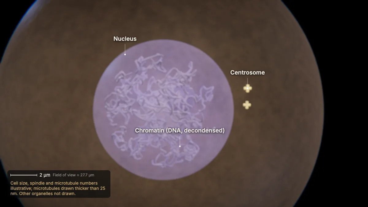
In G1 the cell grows and duplicates organelles; in S it replicates its DNA; in G2 it makes proteins and ATP to prepare for division.
After S phase each chromosome is two identical sister chromatids, joined at a centromere.
The centrosome — the cell's microtubule-organizing center — is duplicated during interphase.
In the model: Cell size, spindle and microtubule numbers illustrative; microtubules drawn thicker than 25 nm. Other organelles not drawn.
3A replicated chromosome
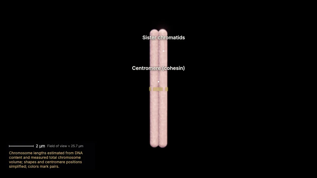
Human body cells have 46 chromosomes — 23 pairs, one set from each parent. After S phase, each is two sister chromatids.
Cohesin proteins hold the sister chromatids together, most tightly at the centromere.
DNA wraps around histones as nucleosomes and packs tighter and tighter: a metaphase chromosome is about 1,400 nm across.
Shown here: chromosome 1, the largest (about 249 million base pairs).
- Chromosomes (human)46 = 23 pairs
- Width≈ 1.4 µm
In the model: Chromosome lengths estimated from DNA content and measured total chromosome volume; shapes and centromere positions simplified; colors mark pairs.
Mitosis
4Prophase
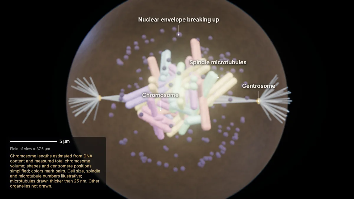
The chromosomes condense and become visible, the nucleolus disappears, and the nuclear envelope starts to break up.
The two centrosomes move toward opposite poles, and the microtubules of the mitotic spindle grow between them.
The Golgi and endoplasmic reticulum break up and scatter toward the cell's edge.
In the model: Chromosome lengths estimated from DNA content and measured total chromosome volume; shapes and centromere positions simplified; colors mark pairs. Cell size, spindle and microtubule numbers illustrative; microtubules drawn thicker than 25 nm. Other organelles not drawn.
5Prometaphase
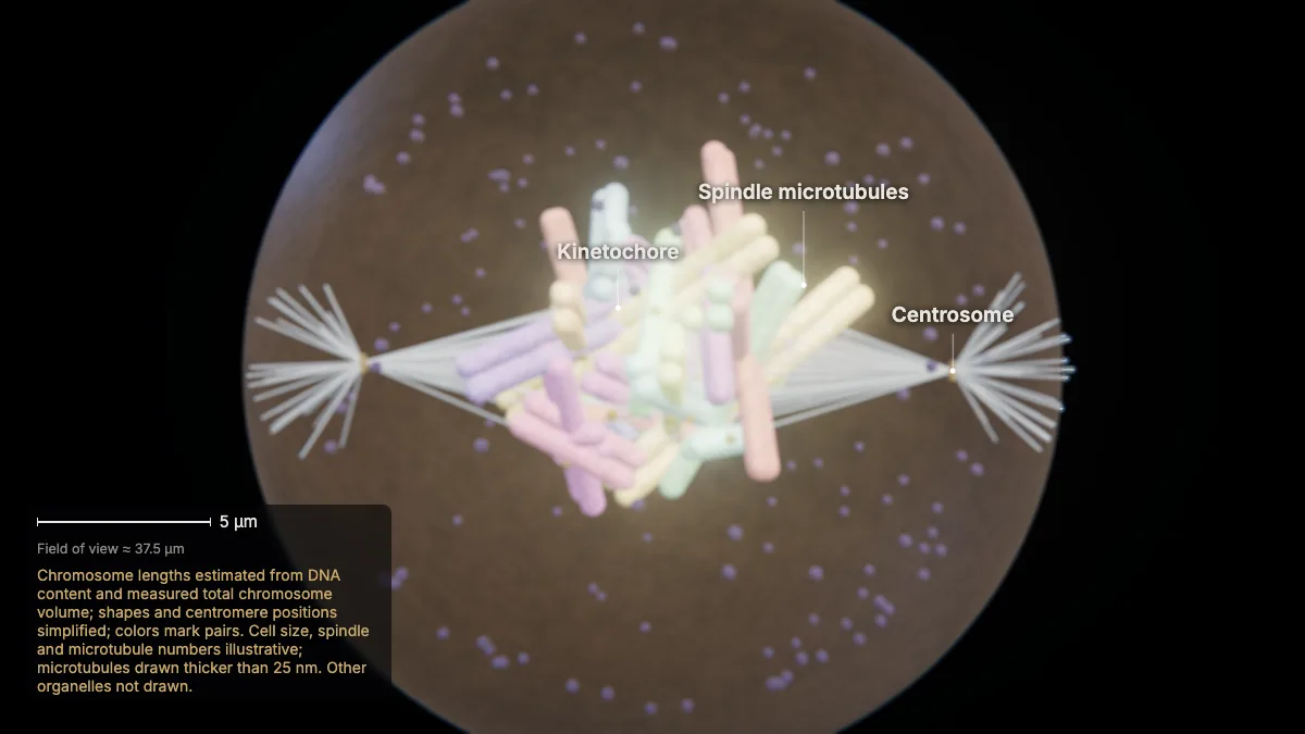
The nuclear envelope breaks into pieces. At its centromere, each sister chromatid forms a kinetochore that binds spindle microtubules.
Spindle microtubules that do not attach to chromosomes overlap between the poles and help lengthen the cell; others radiate from the poles.
In the model: Chromosome lengths estimated from DNA content and measured total chromosome volume; shapes and centromere positions simplified; colors mark pairs. Cell size, spindle and microtubule numbers illustrative; microtubules drawn thicker than 25 nm. Other organelles not drawn.
6Metaphase
All the chromosomes line up in one plane at the cell's equator — the metaphase plate.
Cohesin still holds each pair of sister chromatids together.
A checkpoint waits until every sister chromatid is attached to spindle microtubules before the next step, which cannot be undone.
- Chromosomes at the plate46
In the model: Chromosome lengths estimated from DNA content and measured total chromosome volume; shapes and centromere positions simplified; colors mark pairs. Cell size, spindle and microtubule numbers illustrative; microtubules drawn thicker than 25 nm. Other organelles not drawn.
7Anaphase
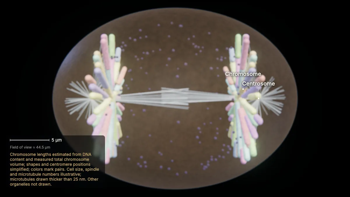
Cohesin breaks down and the sister chromatids separate, pulled to opposite poles. Each chromatid is now its own chromosome.
The cell lengthens as the poles move apart.
- Chromosomes moving to each pole46
In the model: Chromosome lengths estimated from DNA content and measured total chromosome volume; shapes and centromere positions simplified; colors mark pairs. Cell size, spindle and microtubule numbers illustrative; microtubules drawn thicker than 25 nm. Other organelles not drawn.
8Telophase
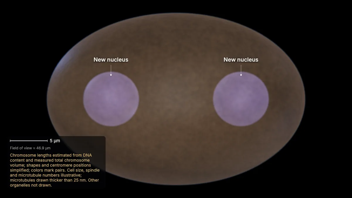
The chromosomes reach the poles and decondense; the spindle breaks down, and a nuclear envelope forms around each set.
In the model: Chromosome lengths estimated from DNA content and measured total chromosome volume; shapes and centromere positions simplified; colors mark pairs. Cell size, spindle and microtubule numbers illustrative; microtubules drawn thicker than 25 nm. Other organelles not drawn.
Cytokinesis
9Cytokinesis
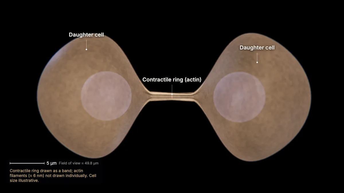
A ring of actin filaments just inside the plasma membrane contracts and pinches the cell in two along a cleavage furrow.
The result: two genetically identical daughter cells, each with a complete genome — 46 chromosomes.
Plant cells divide differently: they build a cell plate between the new nuclei.
In the model: Contractile ring drawn as a band; actin filaments (≈ 6 nm) not drawn individually. Cell size illustrative.
Control and differentiation
10Checkpoints
Checkpoints control the cycle: G1 checks conditions and DNA damage, G2 checks that replication is complete, and the M checkpoint checks spindle attachment.
Cyclins and cyclin-dependent kinases (Cdks) drive the cell through the checkpoints.
When the cycle is disrupted, the result can be cancer — or apoptosis, programmed cell death.
In the model: Chromosome lengths estimated from DNA content and measured total chromosome volume; shapes and centromere positions simplified; colors mark pairs. Cell size, spindle and microtubule numbers illustrative; microtubules drawn thicker than 25 nm. Other organelles not drawn.
11Growth and differentiation
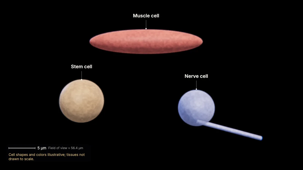
Mitosis drives growth, tissue repair and asexual reproduction; with differentiation, it builds and maintains complex organisms.
All cells of the body, starting with the fertilized egg, contain the same DNA. They specialize by reading only the genes their job needs — switched on or off mainly by transcription factors.
Stem cells are unspecialized cells that can keep dividing as needed.
In the model: Cell shapes and colors illustrative; tissues not drawn to scale.
For teachers
This chapter covers NGSS HS-LS1-4 (the role of mitosis and differentiation in producing and maintaining complex organisms) and AP Biology Topics 4.5 and 4.6 (the cell cycle and its regulation).
- Project the 3D model and go step by step, with labels and an English voice-over you can turn on or off.
- Checkpoints pause the lesson for a question: predict before a step, or check after it.
- Record a lesson video right from the page, or export an MP4.
- Simplifications are stated on screen, and the scale bar is measured from the actual view.
Chapter 1, the tour of the cell, is free for everyone. The Teacher plan adds the next chapters — transcription, translation, protein folding and secretion, cellular respiration, and the cell cycle and mitosis — plus 4K video exports without a watermark: $29 a year during early access (regular price $69).
What the model simplifies
- Chromosome lengths are estimated from each chromosome's DNA content and the measured total volume of metaphase chromosomes; shapes and centromere positions are simplified, and colors only mark pairs.
- Cell size, spindle size and the number of microtubules are illustrative; microtubules (25 nm) are drawn thicker so they are visible.
- Organelles other than the nucleus and centrosomes are not drawn.
- The phases are shown at similar lengths for teaching; interphase really takes most of the cycle.
Sources
- OpenStax, Biology 2e, §10.1–10.3 Cell Reproduction — openstax.org
- OpenStax, Anatomy and Physiology 2e, §3.6 Cellular Differentiation — openstax.org
- College Board, AP Biology Course and Exam Description, Topics 4.5–4.6 — apcentral.collegeboard.org
- NGSS HS-LS1-4 — nextgenscience.org
- Booth et al., 3D-CLEM reveals that a major portion of mitotic chromosomes is not chromatin, Mol Cell 64 (2016) — doi.org
- Ensembl, human genome assembly GRCh38 (chromosome sizes) — ensembl.org