 CYTOTOUR
CytoTour › Photosynthesis
Photosynthesis: the light reactions and the Calvin cycle, in 3D
Photosynthesis uses light energy, carbon dioxide and water to make sugar and releases oxygen: 6 CO₂ + 6 H₂O → C₆H₁₂O₆ + 6 O₂. In plants and algae it happens in chloroplasts. This guide follows the energy of light from the pigments of the thylakoid membrane to the sugar built in the stroma, on real plant protein structures at true size.
The two stages of photosynthesis
| Stage | Where | Takes in | Gives out |
|---|---|---|---|
| Light-dependent reactions | Thylakoid membranes (grana) | Light, H₂O, ADP, NADP⁺ | ATP, NADPH; O₂ released from the split water |
| Calvin cycle (light-independent reactions) | Stroma | CO₂, ATP, NADPH | G3P, used to make glucose and other sugars; ADP and NADP⁺ return |
For one G3P exported (three CO₂ fixed), the Calvin cycle uses 9 ATP and 6 NADPH; one glucose takes six turns.
Step by step, in 3D
These views are Chapter 7 of CytoTour, part of the Teacher plan.
The chloroplast
1Light to sugar
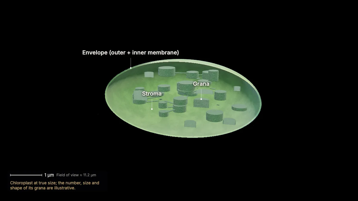
Photosynthesis uses light energy, carbon dioxide and water to make sugar — and releases oxygen.
Overall: 6 CO₂ + 6 H₂O → C₆H₁₂O₆ + 6 O₂, powered by light. Light energy ends up stored as chemical energy in the bonds of sugar.
Plants, algae and cyanobacteria do it; animals, fungi and most other bacteria rely on the sugars it makes. It powers 99 % of Earth's ecosystems.
In plants and algae it happens in chloroplasts — in leaves, mostly in the mesophyll cells.
- Chloroplast≈ 5 µm across, 2.5 µm thick
In the model: Chloroplast at true size; the number, size and shape of its grana are illustrative.
2Inside the chloroplast
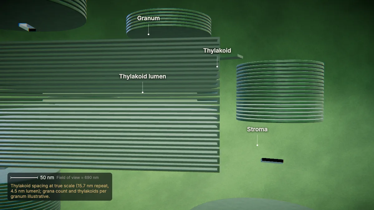
Inside the chloroplast's two envelope membranes are stacks of thylakoids — grana — bathed in a fluid, the stroma.
The space inside each thylakoid is the thylakoid lumen. A mature chloroplast may hold 40–60 grana, each 0.3–0.6 µm across.
The light-dependent reactions happen in the thylakoid membranes; the Calvin cycle happens in the stroma.
Chloroplasts descend from ancient free-living cyanobacteria.
- Granum0.3–0.6 µm across
- Thylakoid repeat15.7 nm
In the model: Thylakoid spacing at true scale (15.7 nm repeat, 4.5 nm lumen); grana count and thylakoids per granum illustrative.
The light-dependent reactions
3The thylakoid membrane
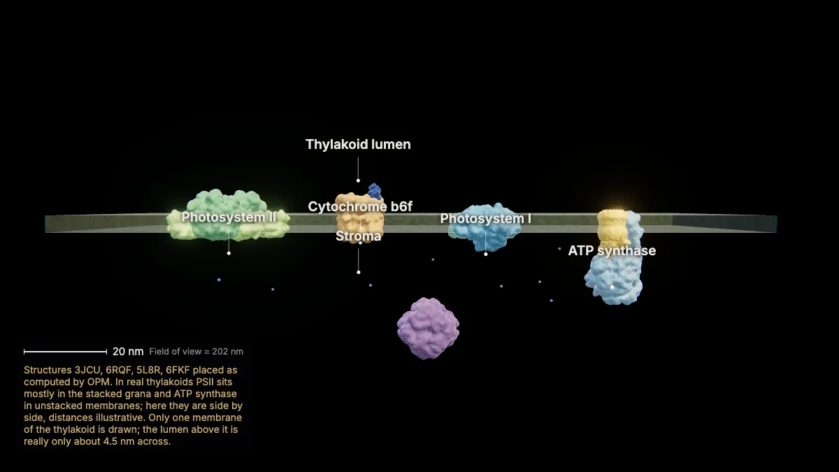
Built into the thylakoid membrane are photosystem II, the cytochrome b6f complex, photosystem I and ATP synthase.
Photosystems II and I are connected by an electron transport chain. They are numbered in the order they were discovered, not the order electrons pass through them.
Each is shown at true size, from spinach and pea structures. The lumen is above the membrane; the stroma below.
- PSII–LHCII≈ 27 nm long
- Membrane≈ 4 nm thick
In the model: Structures 3JCU, 6RQF, 5L8R, 6FKF placed as computed by OPM. In real thylakoids PSII sits mostly in the stacked grana and ATP synthase in unstacked membranes; here they are side by side, distances illustrative. Only one membrane of the thylakoid is drawn; the lumen above it is really only about 4.5 nm across.
4Capturing light
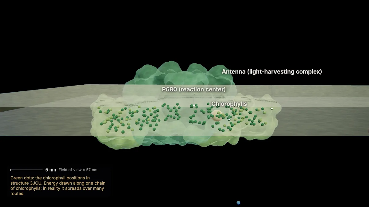
Chlorophyll absorbs blue and red light but not green — that is reflected, so leaves look green.
A photon absorbed anywhere in the antenna excites a chlorophyll. The energy — not an electron — is passed from chlorophyll to chlorophyll until it reaches the reaction center, in about a millionth of a second.
Carotenoids, the other pigments, absorb excess energy and release it as heat.
- Chlorophylls in this PSII–LHCII156 a + 50 b
In the model: Green dots: the chlorophyll positions in structure 3JCU. Energy drawn along one chain of chlorophylls; in reality it spreads over many routes.
5Photosystem II
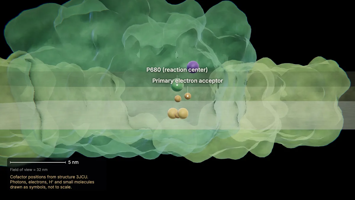
In the reaction center, P680 — a special pair of chlorophyll a — gives up its excited electron to the primary electron acceptor.
This is where light energy becomes an energized electron.
From the acceptor, the electron passes on to the carrier plastoquinone (Pq).
In the model: Cofactor positions from structure 3JCU. Photons, electrons, H⁺ and small molecules drawn as symbols, not to scale.
6Water is split
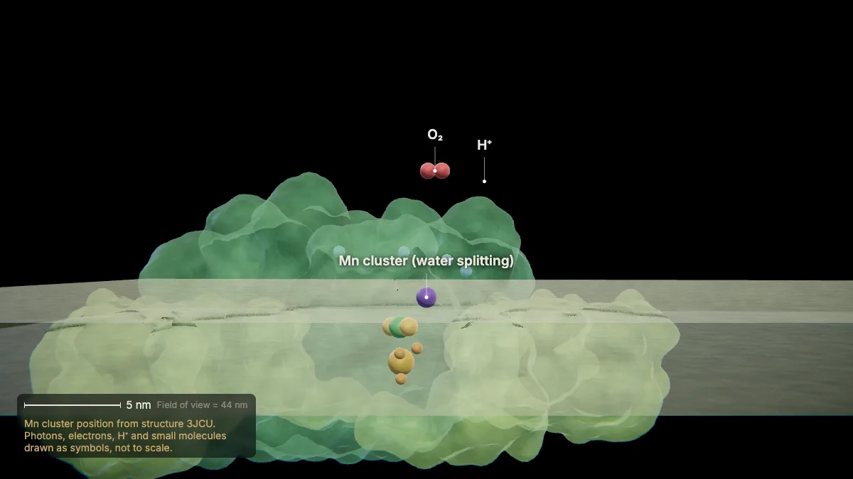
P680's lost electrons are replaced with electrons from water: at a cluster of manganese atoms, water is split.
Splitting two water molecules gives four electrons, four hydrogen ions — left in the lumen — and one O₂.
The oxygen is released as a by-product: its atoms come from water. Some is used by the leaf's own mitochondria; most escapes to the air.
In the model: Mn cluster position from structure 3JCU. Photons, electrons, H⁺ and small molecules drawn as symbols, not to scale.
7The electron transport chain
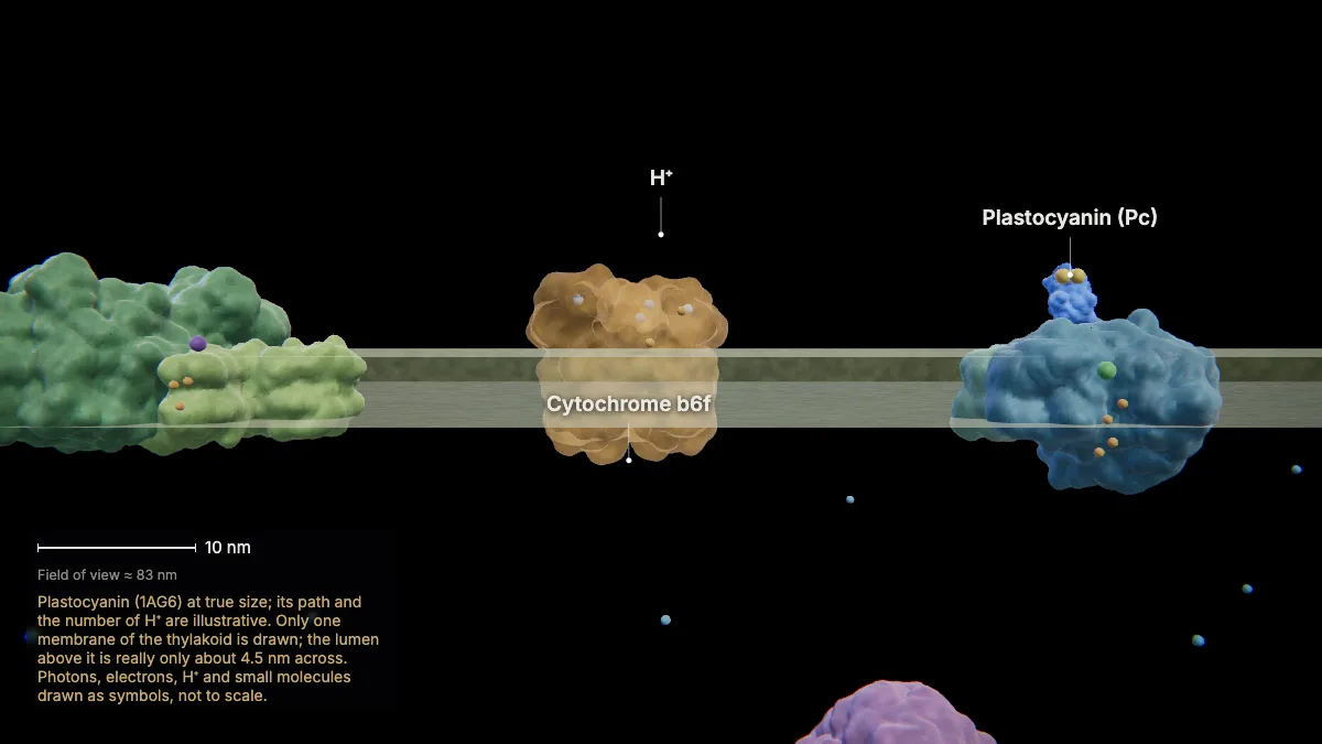
Plastoquinone carries the electrons to the cytochrome b6f complex; the protein plastocyanin then carries them, through the lumen, to photosystem I.
Each hand-over is an oxidation–reduction reaction. The electrons lose energy on the way, and that energy moves hydrogen ions from the stroma into the lumen.
In the model: Plastocyanin (1AG6) at true size; its path and the number of H⁺ are illustrative. Only one membrane of the thylakoid is drawn; the lumen above it is really only about 4.5 nm across. Photons, electrons, H⁺ and small molecules drawn as symbols, not to scale.
8Photosystem I and NADPH
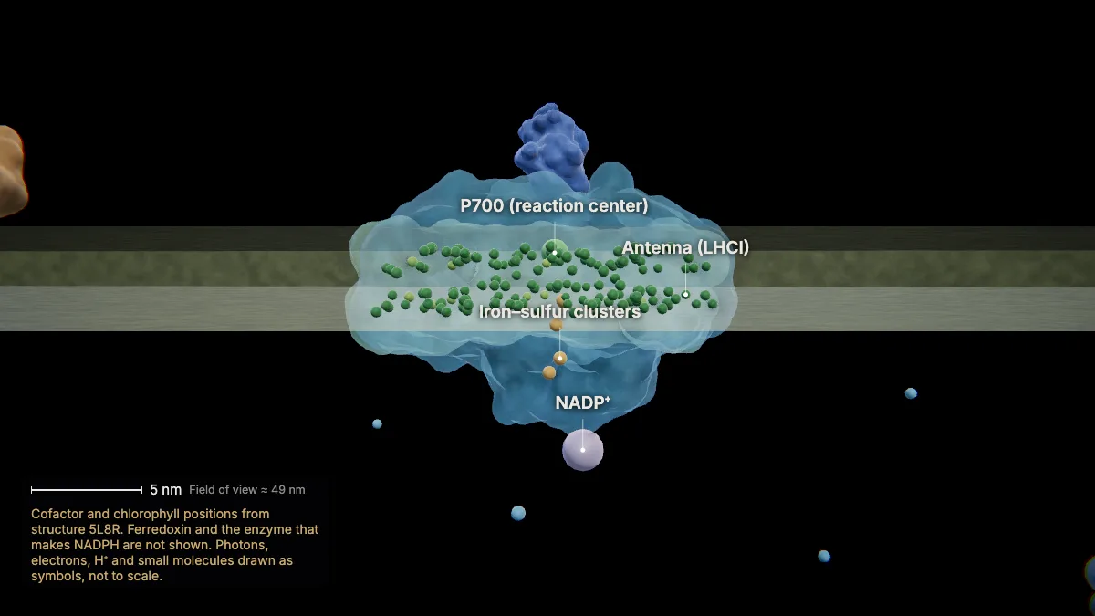
The electrons reach photosystem I with less energy. Another photon, passed to its reaction center P700, energizes them again.
Photosystem I hands the electrons to NADP⁺ in the stroma, reducing it to NADPH.
So photosystem II captures the energy that builds the proton gradient used for ATP; photosystem I, the energy that makes NADPH.
In the model: Cofactor and chlorophyll positions from structure 5L8R. Ferredoxin and the enzyme that makes NADPH are not shown. Photons, electrons, H⁺ and small molecules drawn as symbols, not to scale.
The proton gradient and ATP synthase
9A proton gradient
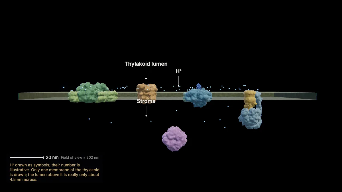
Hydrogen ions build up inside the thylakoid: high in the lumen, low in the stroma — an electrochemical gradient across the membrane.
Protons from splitting water add to the lumen, and making NADPH removes protons from the stroma. The lumen ends up at low pH, the stroma at high pH.
In the model: H⁺ drawn as symbols; their number is illustrative. Only one membrane of the thylakoid is drawn; the lumen above it is really only about 4.5 nm across.
10Chloroplast ATP synthase
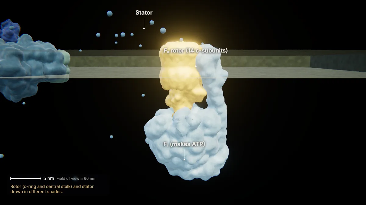
Protons flow back to the stroma through ATP synthase. Its F₁ head sticks into the stroma; F₀ sits in the membrane.
This spinach enzyme has 26 protein subunits. Its rotor ring has 14 c-subunits — the human mitochondrial ring in chapter 5 has 8.
In the thylakoid, ATP synthase sits in the unstacked membranes.
- c-ring14 subunits
- Length (PDB 6FKF)≈ 23 nm
In the model: Rotor (c-ring and central stalk) and stator drawn in different shades.
11Photophosphorylation
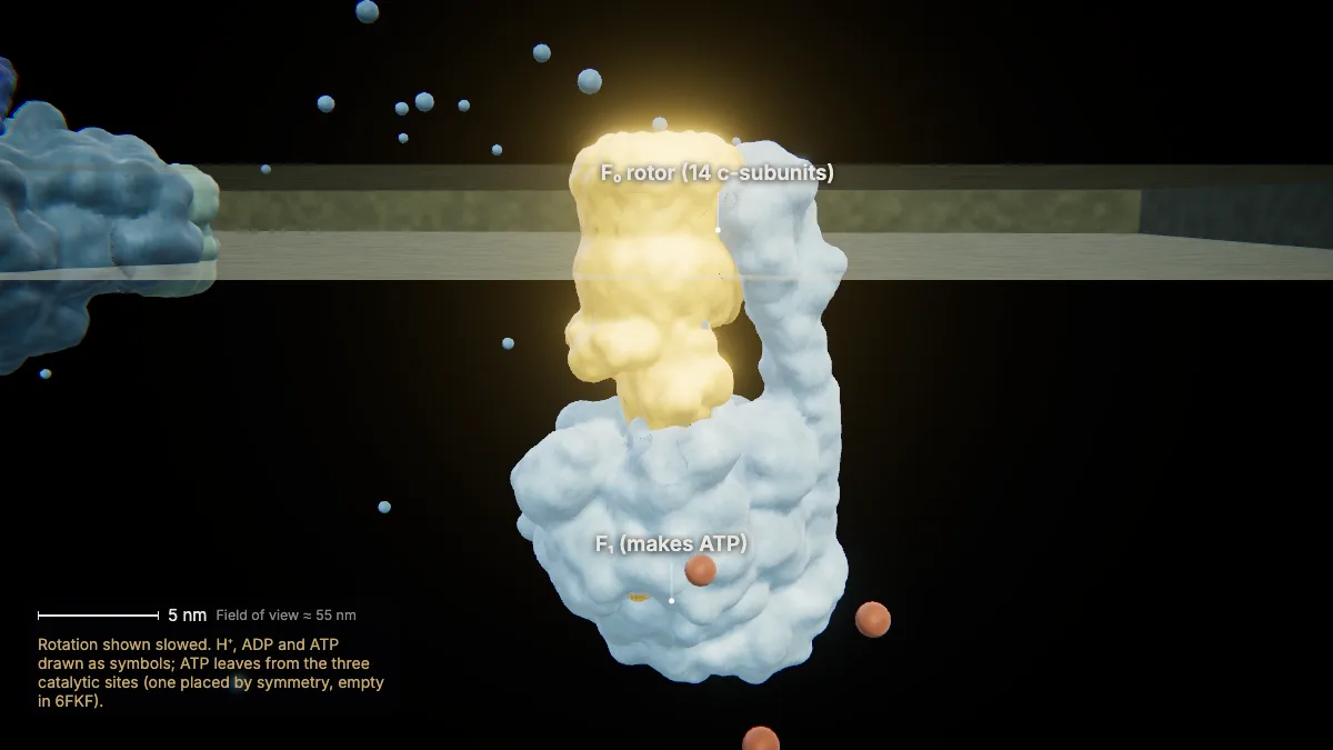
Protons flowing back through F₀ turn the rotor, and F₁ makes ATP from ADP and phosphate — photophosphorylation.
This is chemiosmosis, as in mitochondria. The chloroplast enzyme uses about 4.7 protons for each ATP.
In the model: Rotation shown slowed. H⁺, ADP and ATP drawn as symbols; ATP leaves from the three catalytic sites (one placed by symmetry, empty in 6FKF).
The Calvin cycle
12Fixing carbon
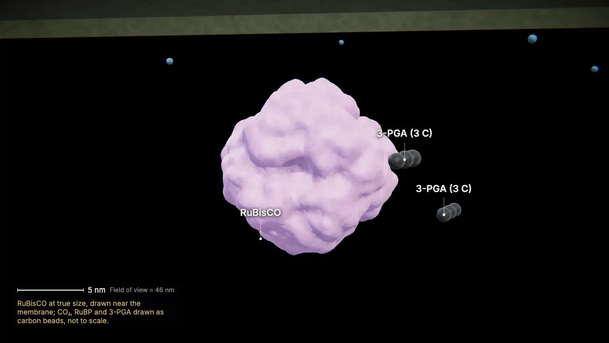
In the stroma, the enzyme RuBisCO joins CO₂ to RuBP, a five-carbon molecule: carbon fixation, from inorganic to organic.
Each CO₂ and RuBP give two molecules of 3-PGA, with three carbons each.
This is the first stage of the Calvin cycle, which runs on the ATP and NADPH from the light reactions.
- RuBisCO (PDB 8RUC)8 large + 8 small subunits
- Size≈ 13 nm
In the model: RuBisCO at true size, drawn near the membrane; CO₂, RuBP and 3-PGA drawn as carbon beads, not to scale.
13The Calvin cycle
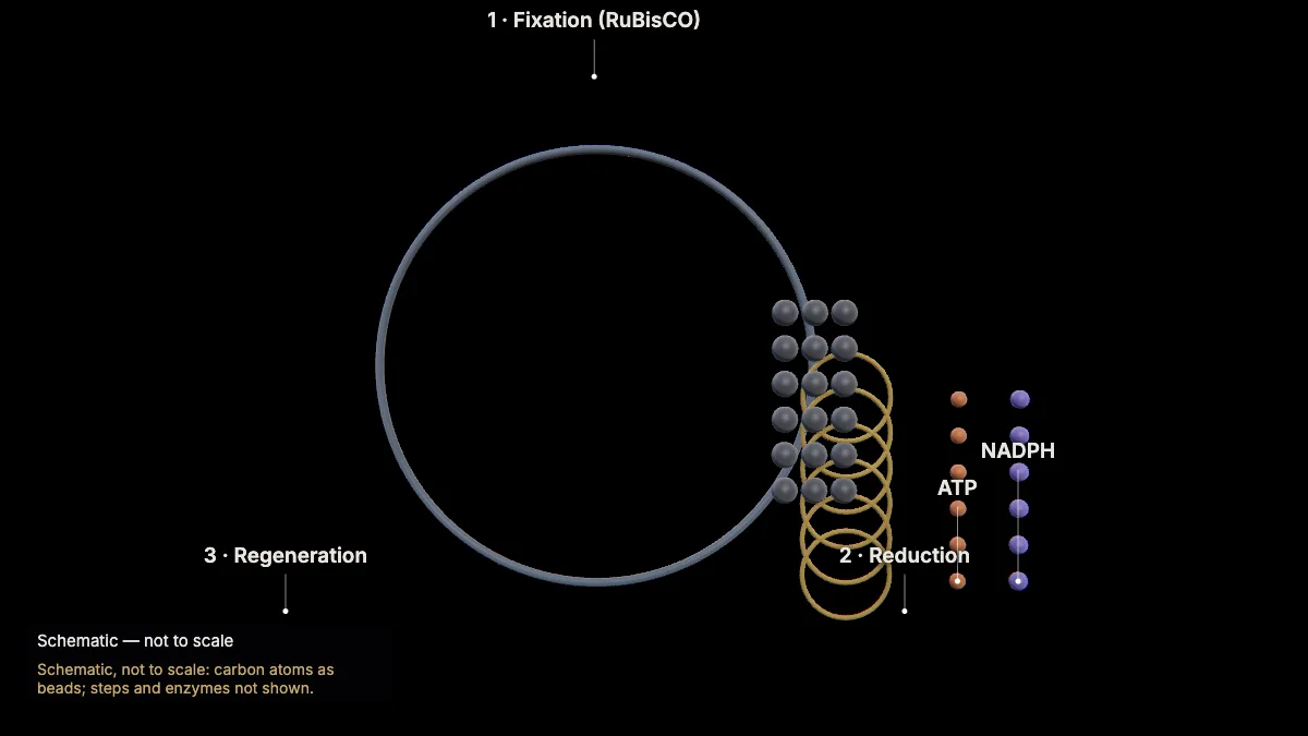
Three turns fix three CO₂. ATP and NADPH reduce the 3-PGA to G3P; one G3P leaves, and the other five rebuild three RuBP.
Fixation: 3 CO₂ + 3 RuBP → 6 3-PGA — 3 + 15 = 18 carbons, all kept. Reduction uses 6 ATP and 6 NADPH; regeneration uses 3 more ATP.
Two exported G3P make one glucose, so six turns are needed per glucose. The ADP and NADP⁺ return to the light reactions.
It was once called the "dark reactions", but it needs the light reactions' products, and some of its enzymes are switched on by light.
- For one G3P3 CO₂ · 9 ATP · 6 NADPH
- Turns per glucose6
In the model: Schematic, not to scale: carbon atoms as beads; steps and enzymes not shown.
Photosynthesis and cellular respiration
14Photosynthesis and respiration
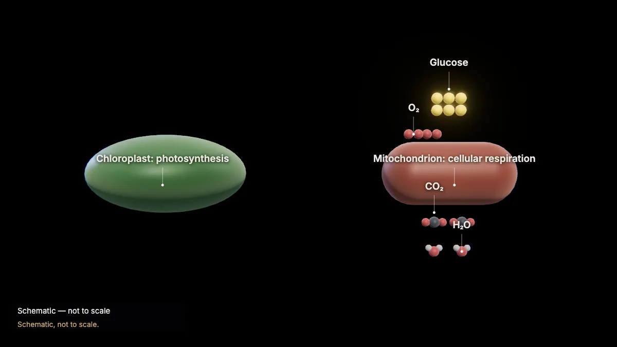
Photosynthesis takes in CO₂ and releases O₂; cellular respiration takes in O₂ and releases CO₂. Together they cycle carbon.
Plants do both: their mitochondria break down the sugars their chloroplasts make. Both processes use an electron transport chain.
Photosynthesis first evolved in prokaryotes; cyanobacteria filled Earth's atmosphere with oxygen.
In the model: Schematic, not to scale.
For teachers
This chapter covers NGSS HS-LS1-5 (how photosynthesis transforms light energy into stored chemical energy) and AP Biology Topic 3.4 (photosynthesis).
- Project the 3D model and go step by step, with labels and an English voice-over you can turn on or off.
- Checkpoints pause the lesson for a question: predict before a step, or check after it.
- Record a lesson video right from the page, or export an MP4.
- Simplifications are stated on screen, and the scale bar is measured from the actual view.
Chapter 1, the tour of the cell, is free for everyone. The Teacher plan adds the next chapters — transcription, translation, protein folding and secretion, cellular respiration, the cell cycle and mitosis, and photosynthesis — plus 4K video exports without a watermark: $29 a year during early access (regular price $69).
What the model simplifies
- The chloroplast is at true size; the number, size and shape of its grana are illustrative. Thylakoid spacing is true to scale (15.7 nm repeat, 4.5 nm lumen).
- In the membrane views the protein structures (spinach and pea) are at true size, placed in the membrane as computed by the OPM database. They are shown side by side, though photosystem II sits mostly in the stacked grana and ATP synthase in unstacked membranes; only one membrane of the thylakoid is drawn.
- Photons, electrons, protons and small molecules are drawn as symbols; energy transfer is drawn along one chain of chlorophylls; ferredoxin and the enzyme that makes NADPH are not shown.
- The Calvin cycle is a schematic map with carbon atoms as beads; its steps and enzymes, other than RuBisCO, are not shown.
- ATP synthase is shown turning slowed down.
Sources
- OpenStax, Biology 2e, §8.1–8.3 Photosynthesis — openstax.org
- College Board, AP Biology Course and Exam Description, Topic 3.4 — apcentral.collegeboard.org
- NGSS HS-LS1-5 — nextgenscience.org
- Staehelin, Chloroplast structure, Photosynth Res 76 (2003), via BioNumbers — bionumbers.hms.harvard.edu
- Daum et al., Arrangement of photosystem II and ATP synthase in chloroplast membranes of spinach and pea, Plant Cell 22 (2010) — doi.org
- Hahn et al., Structure, mechanism, and regulation of the chloroplast ATP synthase, Science 360 (2018) — doi.org
- Ermakova et al., J Exp Bot 73 (2022) — chloroplast ATP synthase H⁺/ATP — doi.org
- UniProt P69560 (photosystem II D1) and P00289 (plastocyanin) — uniprot.org
- Protein Data Bank structures 3JCU, 6RQF, 5L8R, 6FKF, 1AG6, 8RUC — rcsb.org
- OPM — Orientations of Proteins in Membranes — opm.phar.umich.edu