 CYTOTOUR
CytoTour › Cell communication
Cell communication: signal transduction, step by step, in 3D
Cells communicate through direct contact or with chemical signals. This guide follows one signal all the way: the hormone epinephrine reaching a muscle cell, binding a G protein-coupled receptor, and — through a G protein, cAMP and a phosphorylation cascade — freeing glucose from glycogen. Every protein is drawn from a real structure, at true size.
Reception, transduction, response
| Stage | In the epinephrine pathway |
|---|---|
| Reception | Epinephrine binds the β2-adrenergic receptor, a G protein-coupled receptor, on the cell surface |
| Transduction: G protein | The receptor changes shape and activates Gs: Gα swaps GDP for GTP and separates from βγ |
| Transduction: second messenger | Gα activates adenylyl cyclase, which makes cAMP from ATP |
| Transduction: kinase cascade | cAMP activates PKA → phosphorylase kinase → glycogen phosphorylase; the signal is amplified |
| Response | Glycogen is broken down to glucose; glycogen synthase is inhibited |
| Termination | GTP → GDP, cAMP → AMP (phosphodiesterase), phosphates removed (phosphatases) |
Step by step, in 3D
These views are Chapter 9 of CytoTour, part of the Teacher plan.
How cells communicate
1How cells communicate
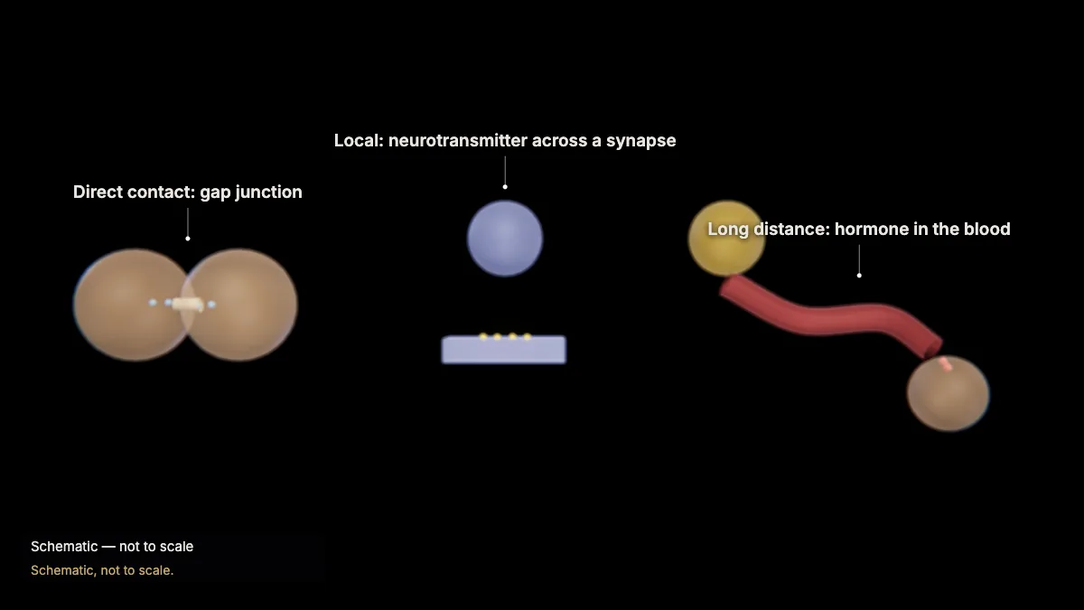
Cells communicate through direct contact or by chemical signals — ligands that bind specific receptor proteins on target cells.
Neighboring animal cells can pass small molecules directly through gap junctions (plants: plasmodesmata).
Local regulators act on nearby cells — for example, neurotransmitters cross a synapse only 20–40 nm wide. Hormones travel in the blood to distant target cells.
Even bacteria signal: in quorum sensing, a signal molecule builds up as the population grows and switches on genes.
In the model: Schematic, not to scale.
2Epinephrine: a hormone
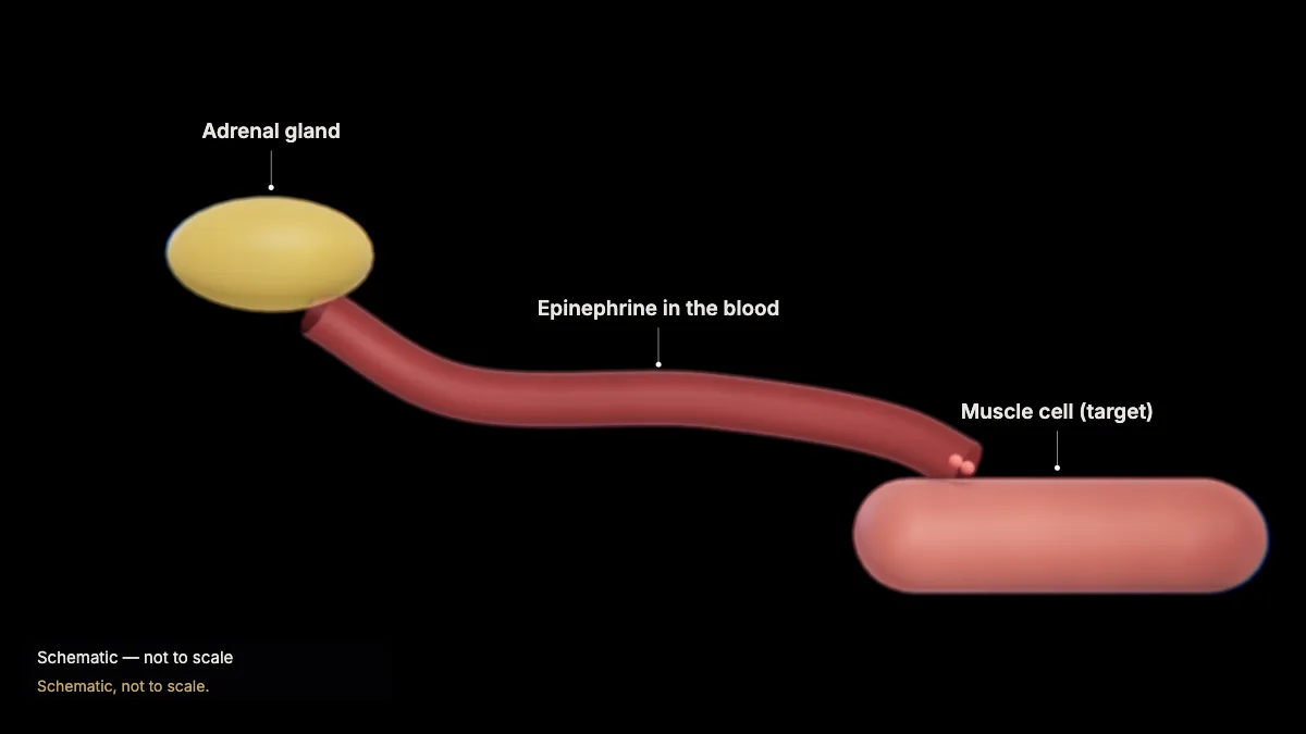
In an emergency, the adrenal glands release the hormone epinephrine (adrenaline), which travels in the blood to muscle cells.
It readies the body for "fight or flight". In a muscle cell it sets off a chain of events that frees glucose from glycogen — followed here molecule by molecule.
In the model: Schematic, not to scale.
From receptor to response
3Reception
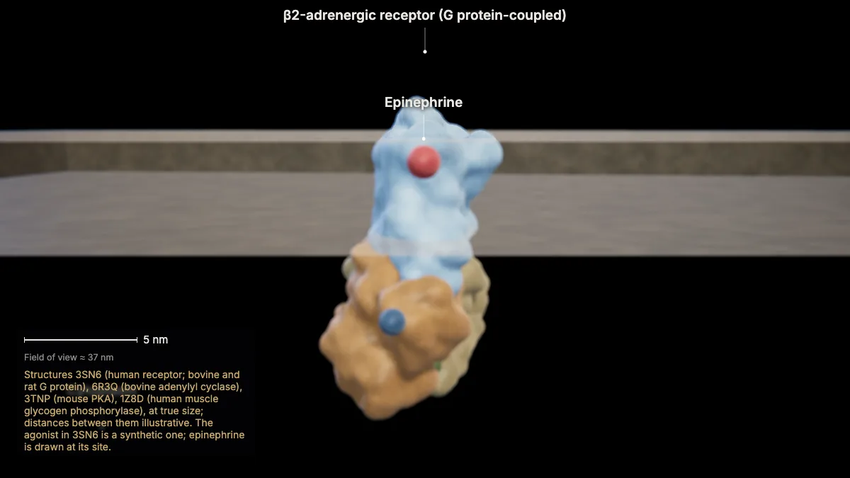
Epinephrine binds its receptor on the muscle cell's surface — the β2-adrenergic receptor, a G protein-coupled receptor.
Its ligand-binding site, open to the outside, recognizes this one kind of molecule; the hormone never has to enter the cell.
Like every G protein-coupled receptor, it crosses the membrane seven times. Signal transduction links this reception to a response inside the cell.
- Receptor (3SN6)≈ 8 nm
In the model: Structures 3SN6 (human receptor; bovine and rat G protein), 6R3Q (bovine adenylyl cyclase), 3TNP (mouse PKA), 1Z8D (human muscle glycogen phosphorylase), at true size; distances between them illustrative. The agonist in 3SN6 is a synthetic one; epinephrine is drawn at its site.
4The G protein switches on
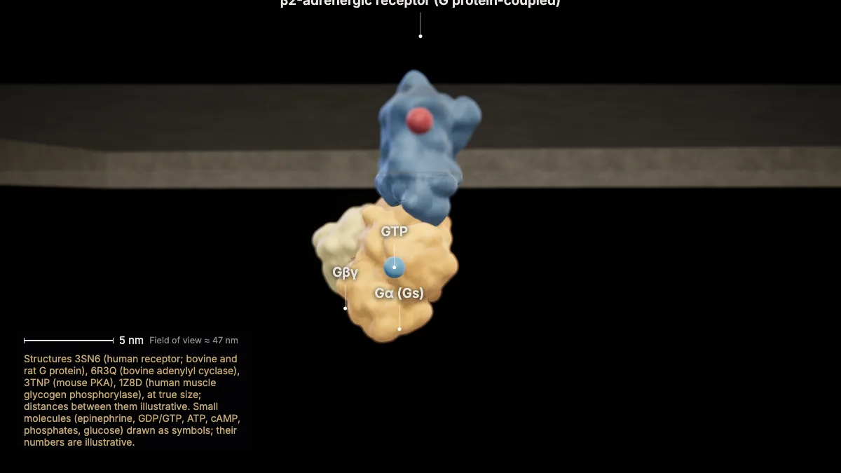
The bound receptor changes shape on the inside and activates its G protein, Gs: Gα drops GDP, picks up GTP and separates from βγ.
The shape change is large: the inner end of one transmembrane helix swings outward by 14 Å.
Gs is the stimulatory G protein for adenylyl cyclase.
In the model: Structures 3SN6 (human receptor; bovine and rat G protein), 6R3Q (bovine adenylyl cyclase), 3TNP (mouse PKA), 1Z8D (human muscle glycogen phosphorylase), at true size; distances between them illustrative. Small molecules (epinephrine, GDP/GTP, ATP, cAMP, phosphates, glucose) drawn as symbols; their numbers are illustrative.
5A second messenger: cAMP
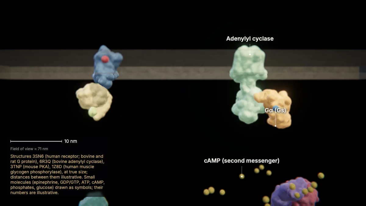
Active Gα binds the membrane enzyme adenylyl cyclase, which turns ATP into cyclic AMP (cAMP).
cAMP is a second messenger: a small molecule that carries the signal through the cytoplasm.
- Adenylyl cyclase 9 (6R3Q)≈ 14 nm
In the model: Structures 3SN6 (human receptor; bovine and rat G protein), 6R3Q (bovine adenylyl cyclase), 3TNP (mouse PKA), 1Z8D (human muscle glycogen phosphorylase), at true size; distances between them illustrative. Small molecules (epinephrine, GDP/GTP, ATP, cAMP, phosphates, glucose) drawn as symbols; their numbers are illustrative.
6Protein kinase A
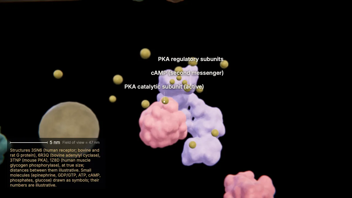
cAMP binds the regulatory subunits of protein kinase A (PKA), and its two catalytic subunits are set free to work.
PKA is a kinase: it moves phosphate groups from ATP onto serines and threonines of its target proteins.
In the model: Structures 3SN6 (human receptor; bovine and rat G protein), 6R3Q (bovine adenylyl cyclase), 3TNP (mouse PKA), 1Z8D (human muscle glycogen phosphorylase), at true size; distances between them illustrative. Small molecules (epinephrine, GDP/GTP, ATP, cAMP, phosphates, glucose) drawn as symbols; their numbers are illustrative.
7A phosphorylation cascade
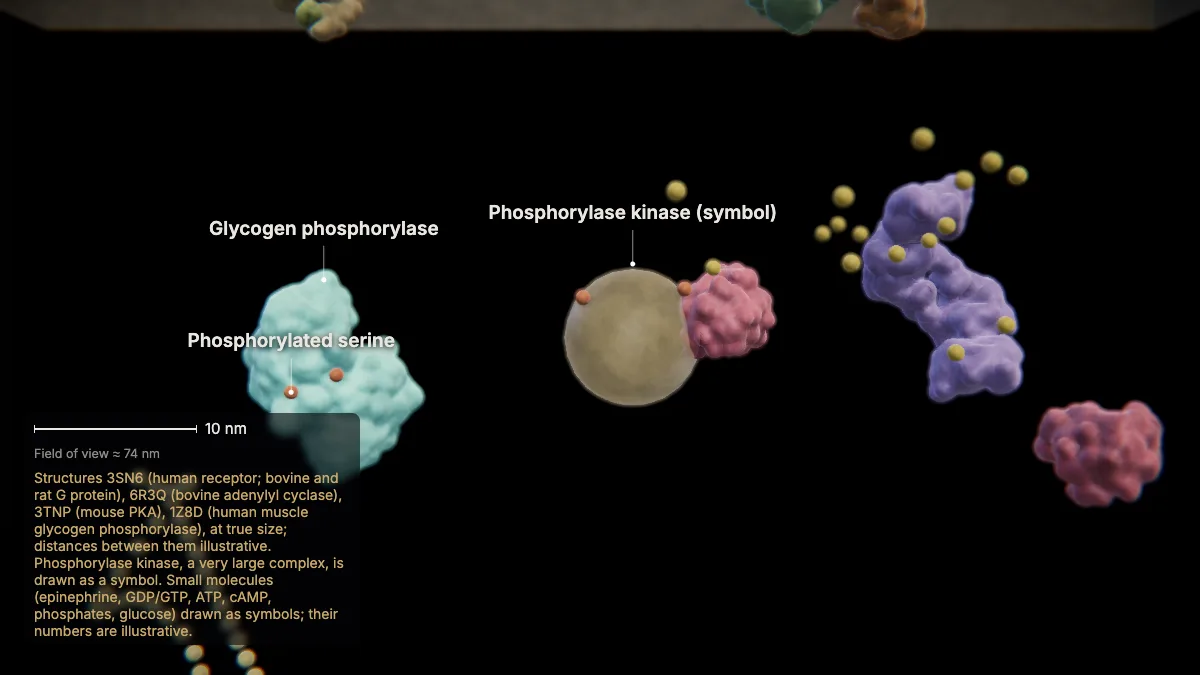
PKA activates phosphorylase kinase, which phosphorylates glycogen phosphorylase — switching it on.
Each step activates many molecules of the next, so the signal is amplified: one hormone at one receptor ends up activating a great many enzymes.
Phosphorylating one serine turns phosphorylase b, inactive, into phosphorylase a, active.
In the model: Structures 3SN6 (human receptor; bovine and rat G protein), 6R3Q (bovine adenylyl cyclase), 3TNP (mouse PKA), 1Z8D (human muscle glycogen phosphorylase), at true size; distances between them illustrative. Phosphorylase kinase, a very large complex, is drawn as a symbol. Small molecules (epinephrine, GDP/GTP, ATP, cAMP, phosphates, glucose) drawn as symbols; their numbers are illustrative.
8The response: glucose
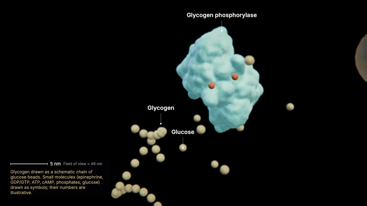
Glycogen phosphorylase breaks glycogen down, freeing glucose for the muscle cell to use.
At the same time PKA phosphorylates glycogen synthase, inhibiting it, so the glucose is not turned straight back into glycogen.
In the model: Glycogen drawn as a schematic chain of glucose beads. Small molecules (epinephrine, GDP/GTP, ATP, cAMP, phosphates, glucose) drawn as symbols; their numbers are illustrative.
9Switching the signal off
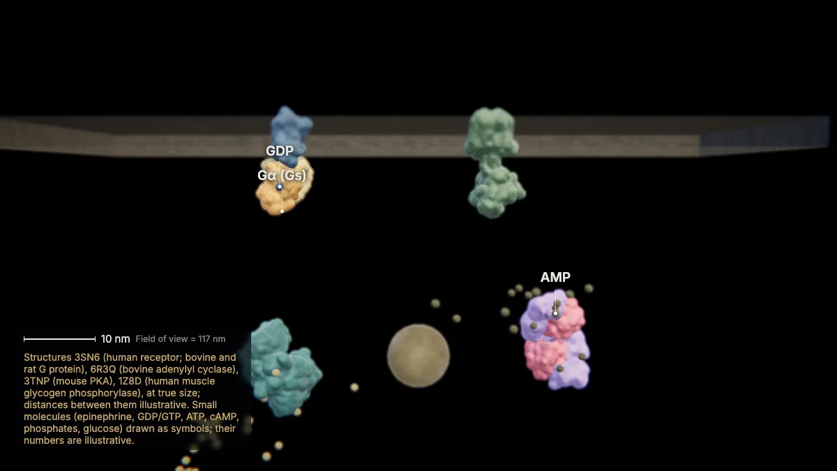
The signal must also stop: Gα turns its GTP into GDP, phosphodiesterase turns cAMP into AMP, and phosphatases remove the phosphates.
Without a fresh signal, the pathway returns to rest.
In the model: Structures 3SN6 (human receptor; bovine and rat G protein), 6R3Q (bovine adenylyl cyclase), 3TNP (mouse PKA), 1Z8D (human muscle glycogen phosphorylase), at true size; distances between them illustrative. Small molecules (epinephrine, GDP/GTP, ATP, cAMP, phosphates, glucose) drawn as symbols; their numbers are illustrative.
Other receptors and responses
10Other kinds of receptors
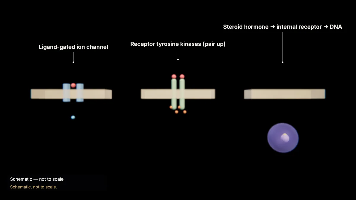
Not all receptors use G proteins. Ligand-gated channels open for ions; receptor tyrosine kinases pair up and phosphorylate each other.
Small hydrophobic hormones such as estrogen, testosterone and thyroid hormones cross the membrane and bind receptors inside the cell, which act on DNA.
In the model: Schematic, not to scale.
11Many possible responses
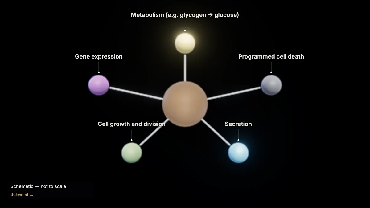
Depending on the cell and the signal, the response can be a change in metabolism, gene expression, growth, secretion — or programmed cell death.
In the model: Schematic.
12When a pathway goes wrong
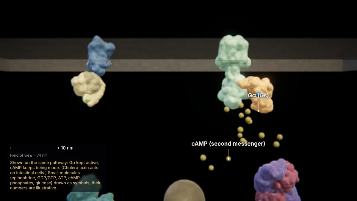
A change in any part of a pathway can change the response. Cholera toxin, for example, keeps a G protein permanently switched on.
Mutations can do the same: about 30 % of cancer cells carry a mutation in RAS, another G protein, that leads to uncontrolled growth.
Chemicals that act on any component — drugs, toxins — can activate or inhibit a pathway.
In the model: Shown on the same pathway: Gα kept active, cAMP keeps being made. (Cholera toxin acts on intestinal cells.) Small molecules (epinephrine, GDP/GTP, ATP, cAMP, phosphates, glucose) drawn as symbols; their numbers are illustrative.
Feedback and homeostasis
13Negative feedback
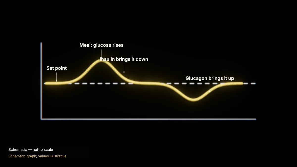
Negative feedback pushes a variable back toward its set point. After a meal, insulin brings blood glucose down; between meals, glucagon brings it up.
Glucagon raises blood glucose by stimulating the breakdown of glycogen. Negative feedback keeps the internal environment steady — homeostasis.
In the model: Schematic graph; values illustrative.
14Positive feedback
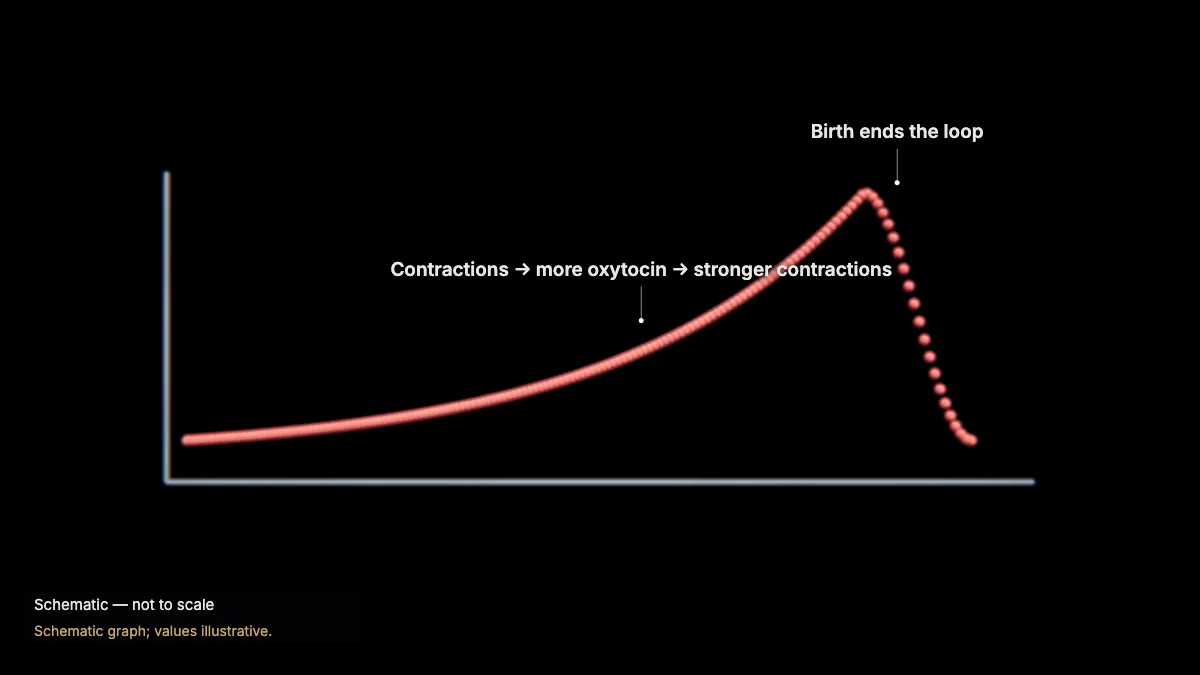
Positive feedback amplifies a change. In childbirth, oxytocin causes contractions, which cause more oxytocin — until the baby is born.
It moves the variable further from where it started, and it ends when the process is complete.
In the model: Schematic graph; values illustrative.
For teachers
This chapter covers AP Biology Topics 4.1–4.4 (cell communication, signal transduction, signal transduction pathways and feedback), using the CED's own example of epinephrine stimulating glycogen breakdown.
- Project the 3D model and go step by step, with labels and an English voice-over you can turn on or off.
- Checkpoints pause the lesson for a question: predict before a step, or check after it.
- Record a lesson video right from the page, or export an MP4.
- Simplifications are stated on screen, and the scale bar is measured from the actual view.
Chapter 1, the tour of the cell, is free for everyone, and a free account also opens cellular respiration and photosynthesis. The Teacher plan adds the other chapters — transcription, translation, protein folding and secretion, the cell cycle and mitosis, meiosis, cell communication, and enzymes — plus worksheet answer keys and 4K video exports without a watermark: $29 a year during early access (regular price $69).
What the model simplifies
- The protein structures are at true size and the membrane proteins are placed as computed by the OPM database; some come from other mammals (bovine, rat, mouse), as stated on screen. Distances between them are illustrative.
- Phosphorylase kinase, a very large complex, is drawn as a symbol; small molecules (epinephrine, GDP/GTP, ATP, cAMP, phosphates, glucose) are symbols and their numbers are illustrative.
- Glycogen is drawn as a schematic chain of glucose beads; the agonist in the receptor structure is a synthetic one, and epinephrine is drawn at its site.
- The kinds of signaling, other receptors, responses and feedback graphs are schematic.
Sources
- OpenStax, Biology 2e, §9.1–9.4 Cell Communication — openstax.org
- OpenStax, Biology 2e, §33.3 Homeostasis and §37.3 Regulation of Body Processes — openstax.org
- College Board, AP Biology Course and Exam Description, Topics 4.1–4.4 — apcentral.collegeboard.org
- Rasmussen et al., Crystal structure of the β2 adrenergic receptor–Gs protein complex, Nature 477 (2011) — doi.org
- Qi et al., The structure of a membrane adenylyl cyclase bound to an activated stimulatory G protein, Science 364 (2019) — doi.org
- Zhang et al., Structure and allostery of the PKA RIIβ tetrameric holoenzyme, Science 335 (2012) — doi.org
- Protein Data Bank structures 3SN6, 6R3Q, 3TNP, 1Z8D; UniProt P11217 — rcsb.org
- OPM — Orientations of Proteins in Membranes — opm.phar.umich.edu