 CYTOTOUR
CytoTour › Meiosis
Meiosis: the stages, crossing over and independent assortment, in 3D
Meiosis makes the gametes for sexual reproduction: one round of chromosome replication, then two divisions, so each gamete gets one set of chromosomes instead of two. This guide follows a human cell — all 46 of its chromosomes, maternal in pink and paternal in blue — to four genetically different haploid cells.
The stages at a glance
| Stage | What happens |
|---|---|
| Prophase I | In prophase I the homologous chromosomes find each other and pair up along their whole length — synapsis. |
| Prophase I: crossing over | Nonsister chromatids of a homologous pair exchange matching segments of DNA — crossing over. |
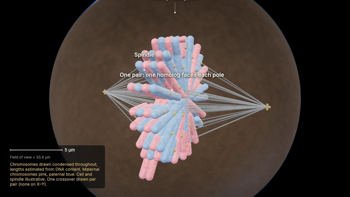
| Metaphase I | The 23 homologous pairs line up at the metaphase plate — as pairs, one homolog facing each pole. |
| Anaphase I | The homologous chromosomes separate and are pulled to opposite poles. Sister chromatids stay together. |
| Telophase I | The cell divides in two. Each new cell is haploid — 23 chromosomes, each still made of two sister chromatids. |
| Metaphase II | Meiosis II works like mitosis: in each cell a new spindle forms and the 23 chromosomes line up at the metaphase plate. |
| Anaphase II | The proteins holding the sisters together at the centromere break down, and the sister chromatids are pulled to opposite poles. |
| Telophase II | Telophase II and cytokinesis leave four haploid cells, each with 23 single-chromatid chromosomes — and no two alike. |
Meiosis vs mitosis: Both use a spindle to move chromosomes, but mitosis makes two identical cells with the parent's number, and meiosis four different cells with half.
Step by step, in 3D
These views are Chapter 8 of CytoTour, part of the Teacher plan.
Why meiosis
1Why meiosis?
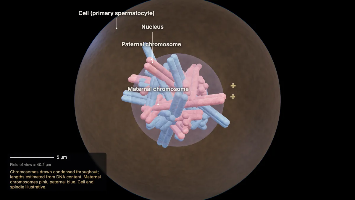
Sexual reproduction joins two gametes — egg and sperm — each with one set of chromosomes. Meiosis makes those gametes, with half the chromosomes.
One set of chromosomes is haploid; two sets are diploid. Without halving, the number of sets would double with every fertilization.
This human cell, about to begin meiosis, has 46 chromosomes: one set of 23 from the mother's egg and one set of 23 from the father's sperm.
Meiosis takes one round of chromosome replication, then two divisions — meiosis I and meiosis II.
- This cell46 chromosomes (2n)
In the model: Chromosomes drawn condensed throughout; lengths estimated from DNA content. Maternal chromosomes pink, paternal blue. Cell and spindle illustrative.
2Homologous chromosomes
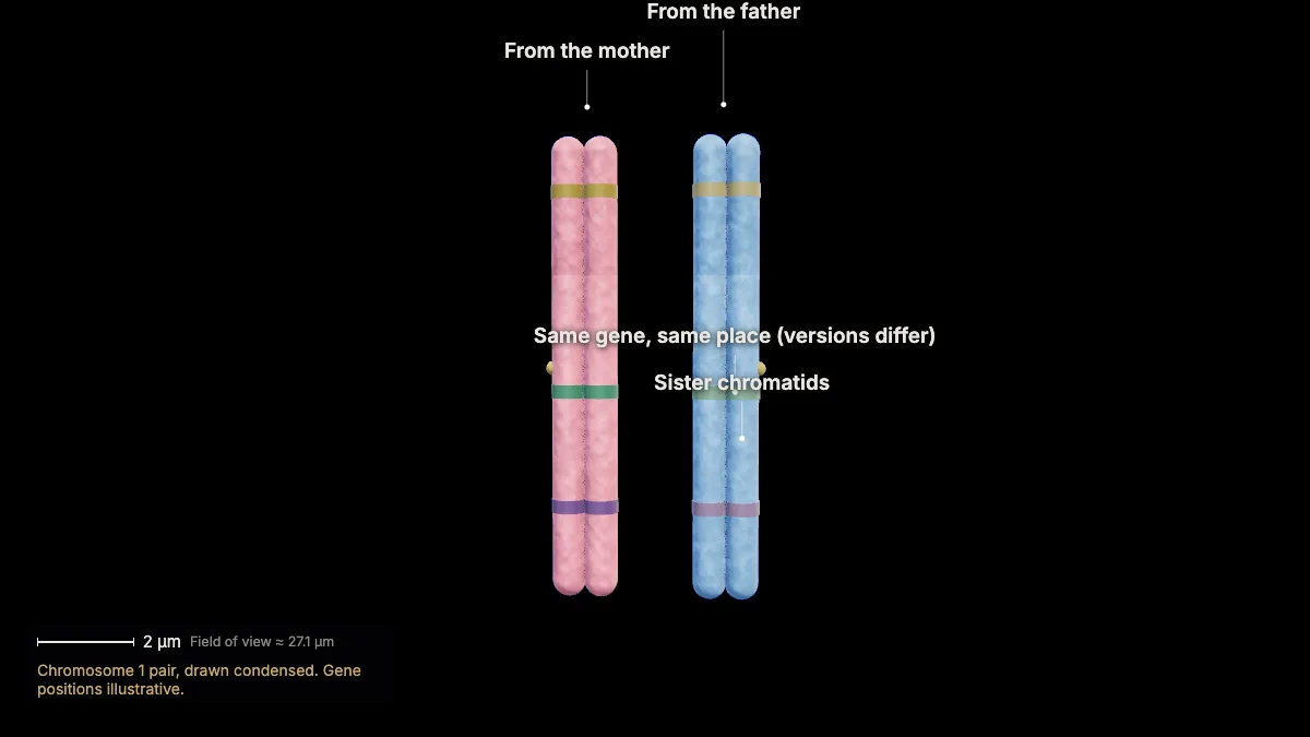
Each chromosome has a partner — its homolog — with the same genes at the same locations: one from the mother, one from the father.
The two homologs are not identical: they can carry different versions of the same genes.
Before meiosis, in S phase, each chromosome was replicated into two sister chromatids, held together at the centromere by cohesin.
- Chromosome 1≈ 9.5 µm long (drawn)
In the model: Chromosome 1 pair, drawn condensed. Gene positions illustrative.
Meiosis I
3Prophase I: homologs pair
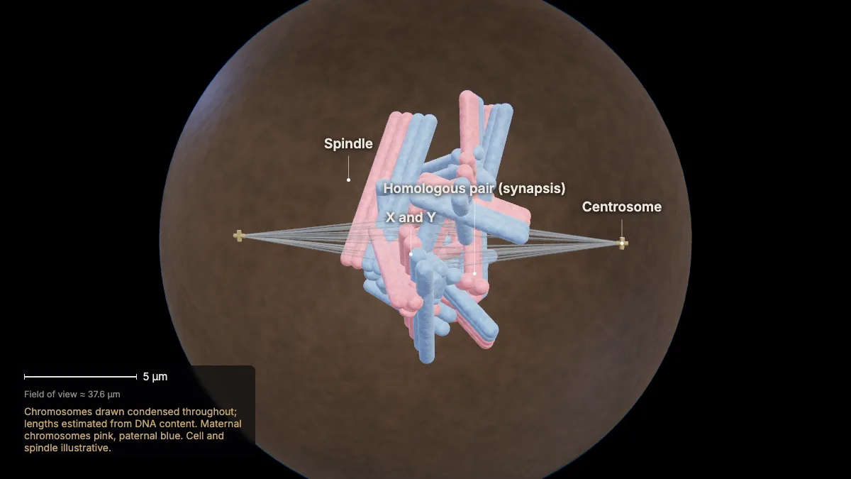
In prophase I the homologous chromosomes find each other and pair up along their whole length — synapsis.
A protein lattice, the synaptonemal complex, holds each pair tightly together. Homologs never pair like this in mitosis.
Meanwhile the chromosomes condense, the centrosomes move to opposite poles as the spindle forms, and the nuclear envelope breaks down.
The X and Y, though mostly different, pair through a small shared region.
In the model: Chromosomes drawn condensed throughout; lengths estimated from DNA content. Maternal chromosomes pink, paternal blue. Cell and spindle illustrative.
4Crossing over
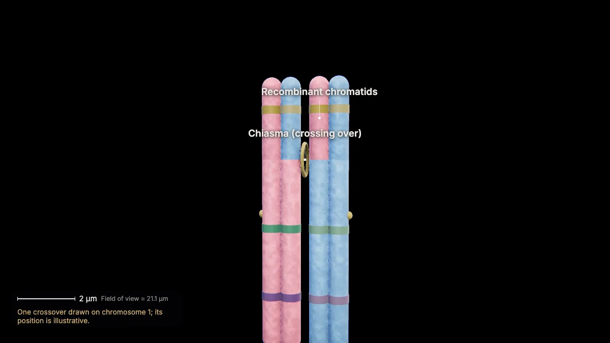
Nonsister chromatids of a homologous pair exchange matching segments of DNA — crossing over.
The exchange points show up as chiasmata. Every pair needs at least one to separate properly; there can be as many as 25.
The recombinant chromatids now carry combinations of maternal and paternal genes that did not exist before.
Held together only at the chiasmata, each pair of four chromatids is called a tetrad.
In the model: One crossover drawn on chromosome 1; its position is illustrative.
5Metaphase I
The 23 homologous pairs line up at the metaphase plate — as pairs, one homolog facing each pole.
Microtubules from opposite poles attach to the two homologs of each pair.
- At the plate23 pairs
In the model: Chromosomes drawn condensed throughout; lengths estimated from DNA content. Maternal chromosomes pink, paternal blue. Cell and spindle illustrative. One crossover drawn per pair (none on X–Y).
6Independent assortment
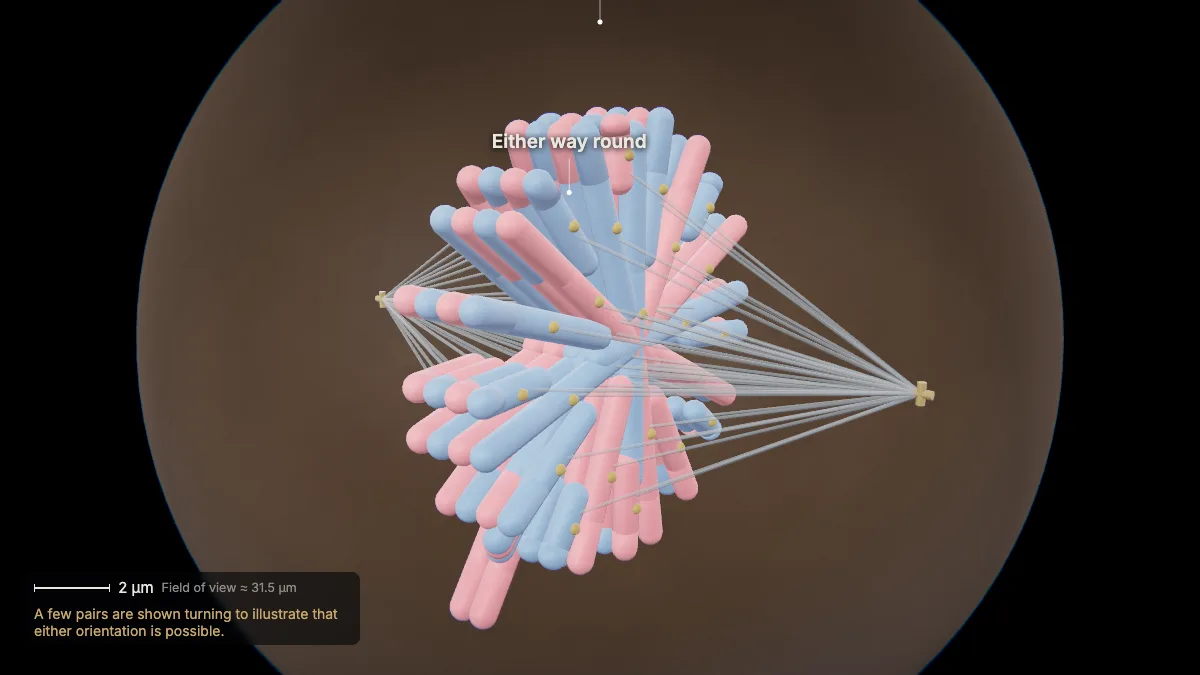
Each pair lines up either way: maternal or paternal homolog toward a pole, independently of every other pair.
With 23 pairs there are 2²³ — over eight million — possible arrangements, before counting crossing over.
- Arrangements2²³ = 8,388,608
In the model: A few pairs are shown turning to illustrate that either orientation is possible.
7Anaphase I
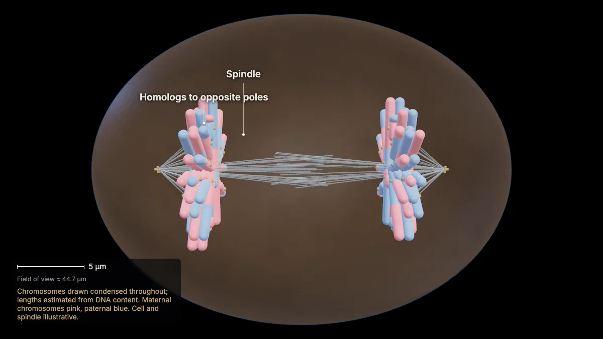
The homologous chromosomes separate and are pulled to opposite poles. Sister chromatids stay together.
The chiasmata are broken; the cohesin at the centromeres still holds each pair of sisters.
In the model: Chromosomes drawn condensed throughout; lengths estimated from DNA content. Maternal chromosomes pink, paternal blue. Cell and spindle illustrative.
8Telophase I: two haploid cells
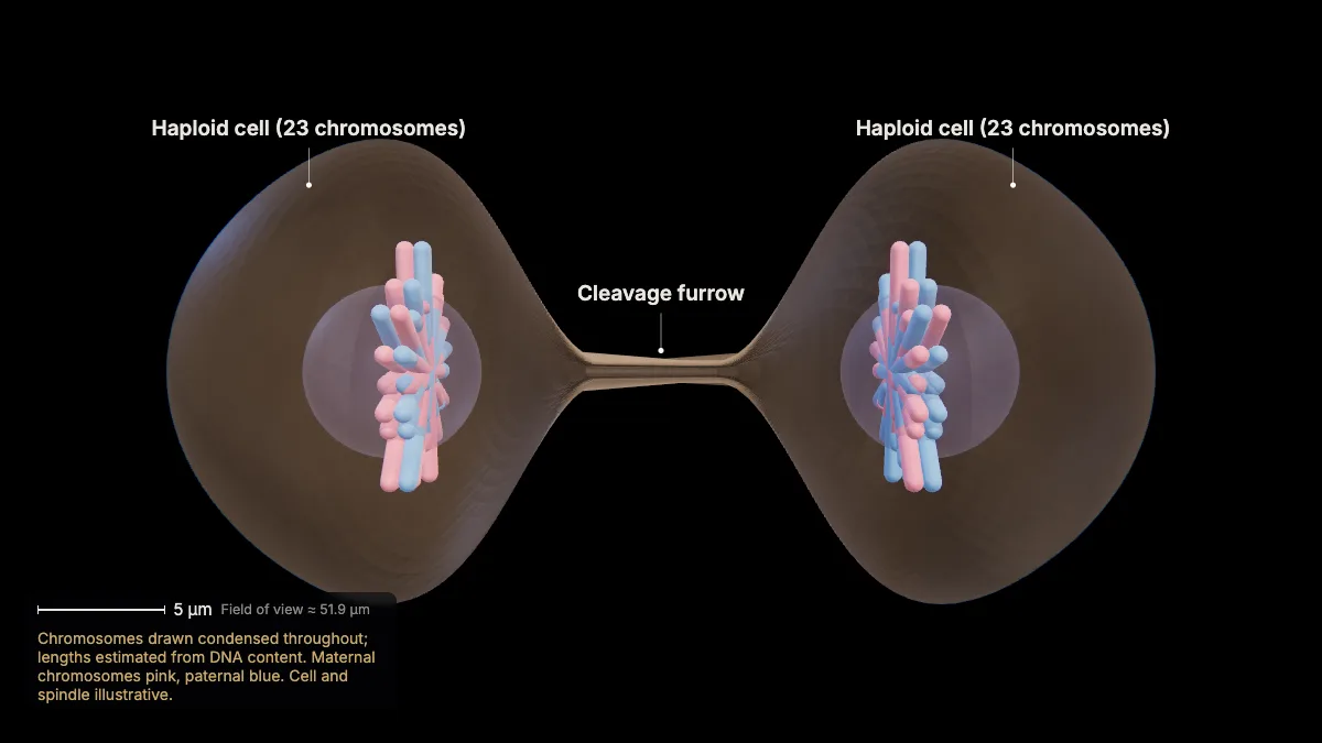
The cell divides in two. Each new cell is haploid — 23 chromosomes, each still made of two sister chromatids.
The spindle breaks down and a cleavage furrow splits the cell. Meiosis I is the reductional division: from two sets to one.
Before the second division, the chromosomes are not replicated again.
- Each cell23 chromosomes (n)
In the model: Chromosomes drawn condensed throughout; lengths estimated from DNA content. Maternal chromosomes pink, paternal blue. Cell and spindle illustrative.
Meiosis II
9Meiosis II: metaphase II
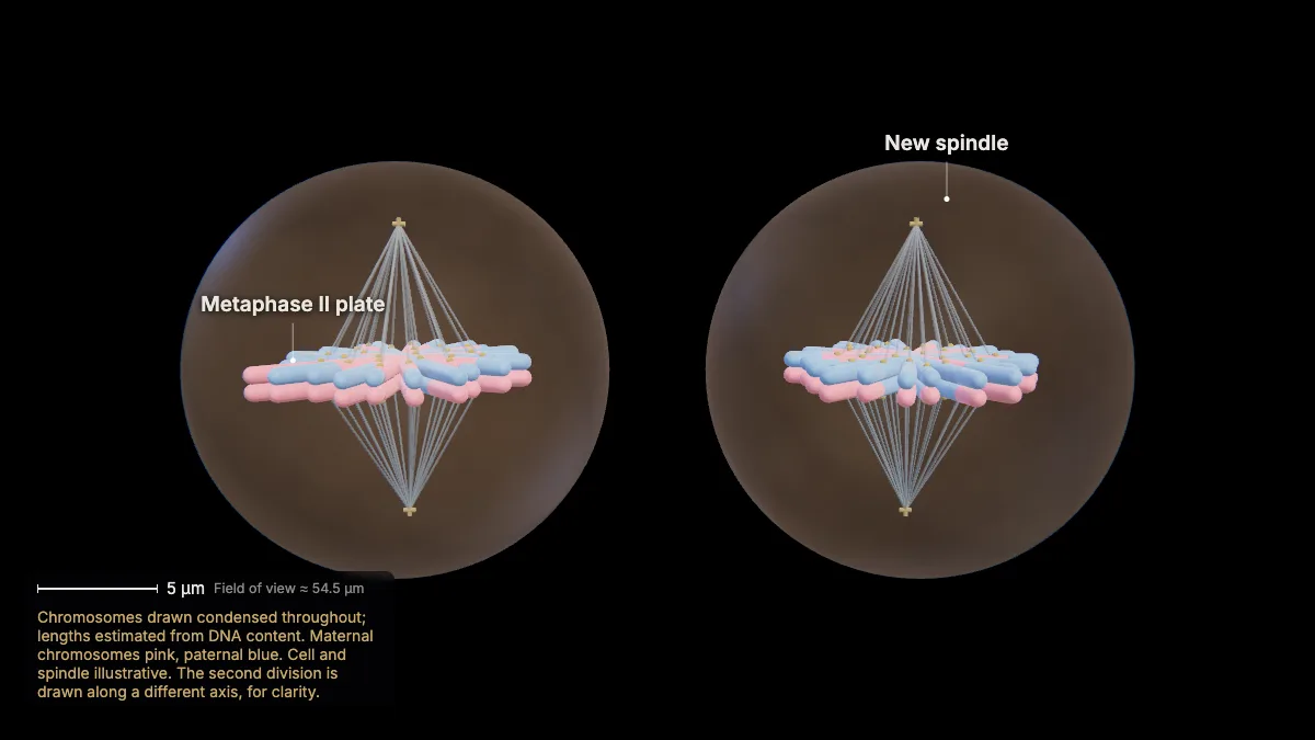
Meiosis II works like mitosis: in each cell a new spindle forms and the 23 chromosomes line up at the metaphase plate.
Now the kinetochore of each sister chromatid is attached to a microtubule from one pole.
In the model: Chromosomes drawn condensed throughout; lengths estimated from DNA content. Maternal chromosomes pink, paternal blue. Cell and spindle illustrative. The second division is drawn along a different axis, for clarity.
10Anaphase II
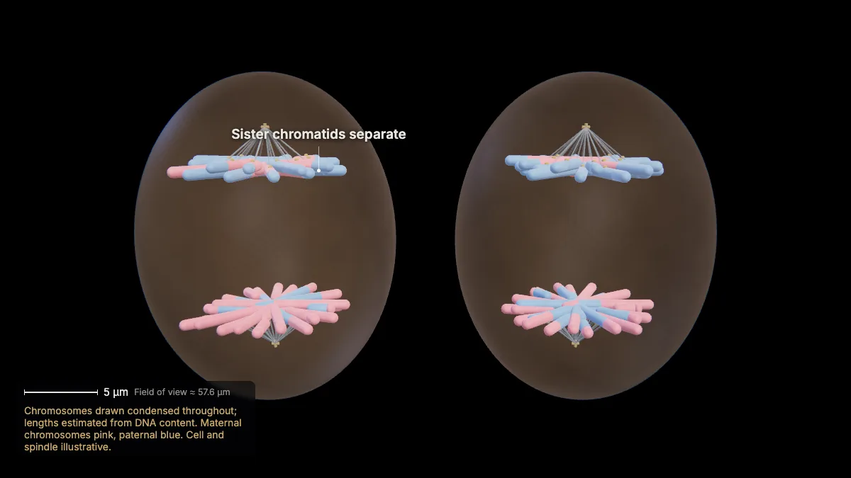
The proteins holding the sisters together at the centromere break down, and the sister chromatids are pulled to opposite poles.
In the model: Chromosomes drawn condensed throughout; lengths estimated from DNA content. Maternal chromosomes pink, paternal blue. Cell and spindle illustrative.
11Four different haploid cells
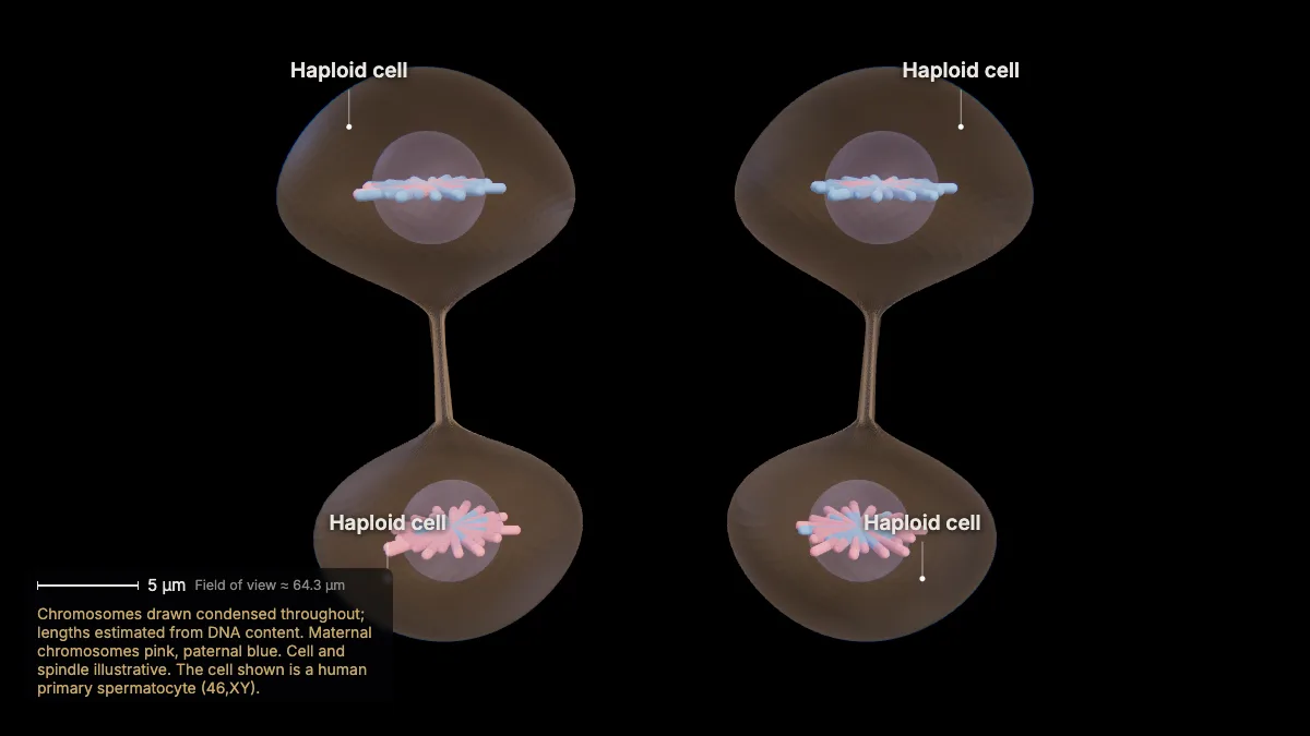
Telophase II and cytokinesis leave four haploid cells, each with 23 single-chromatid chromosomes — and no two alike.
They differ because of crossing over and the random assortment of maternal and paternal homologs.
In males, these four cells become four sperm. In females, one egg is made per meiosis; the extra chromosomes go into polar bodies.
- Each cell23 chromosomes (n)
In the model: Chromosomes drawn condensed throughout; lengths estimated from DNA content. Maternal chromosomes pink, paternal blue. Cell and spindle illustrative. The cell shown is a human primary spermatocyte (46,XY).
Meiosis vs mitosis, nondisjunction and fertilization
12Meiosis vs mitosis
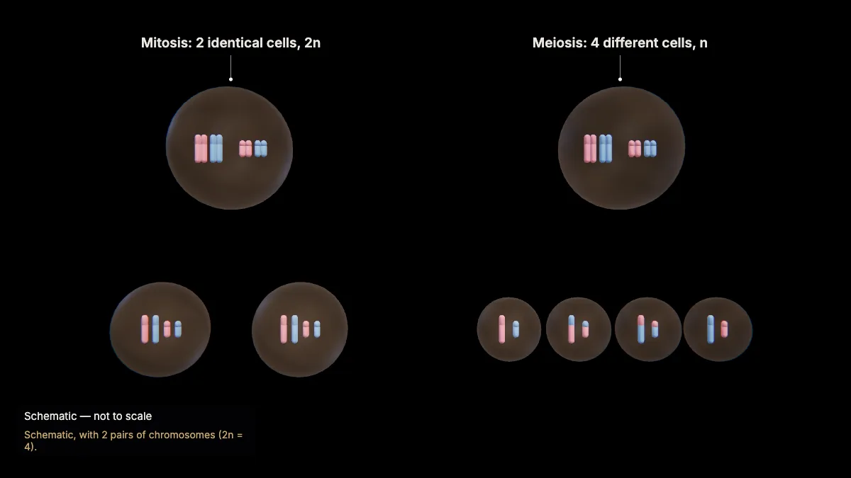
Both use a spindle to move chromosomes, but mitosis makes two identical cells with the parent's number, and meiosis four different cells with half.
Synapsis, crossing over and homologs paired at the plate happen only in meiosis I.
- Mitosis1 division → 2 identical cells (2n)
- Meiosis2 divisions → 4 different cells (n)
In the model: Schematic, with 2 pairs of chromosomes (2n = 4).
13When separation fails
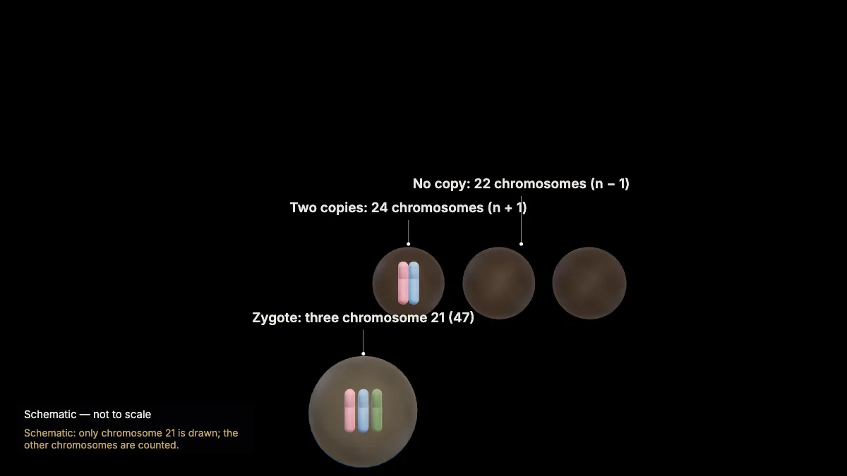
If homologs fail to separate in meiosis I — nondisjunction — two gametes get an extra copy of a chromosome and two get none.
Correct separation gives every gamete one full set. If a gamete with two copies of chromosome 21 joins a normal one, the zygote has three: trisomy 21, which causes Down syndrome.
Nondisjunction in meiosis II gives one gamete too many, one too few and two normal. The risk increases with the parents' age.
- Gametes24, 24, 22, 22 chromosomes
- Zygote47 (three chromosome 21)
In the model: Schematic: only chromosome 21 is drawn; the other chromosomes are counted.
14Fertilization
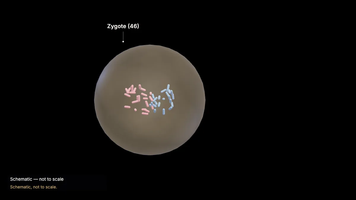
At fertilization a sperm and an egg, each with 23 chromosomes, fuse into a zygote with 46 — a new combination of maternal and paternal chromosomes.
Crossing over, independent assortment and fertilization together make sexual reproduction a source of genetic variation.
- Chromosomes23 + 23 = 46
In the model: Schematic, not to scale.
For teachers
This chapter covers AP Biology Topics 5.1 and 5.2 (meiosis, and meiosis and genetic diversity) and NGSS HS-LS3-2 (inheritable variation from new genetic combinations through meiosis).
- Project the 3D model and go step by step, with labels and an English voice-over you can turn on or off.
- Checkpoints pause the lesson for a question: predict before a step, or check after it.
- Record a lesson video right from the page, or export an MP4.
- Simplifications are stated on screen, and the scale bar is measured from the actual view.
Chapter 1, the tour of the cell, is free for everyone, and a free account also opens cellular respiration and photosynthesis. The Teacher plan adds the other chapters — transcription, translation, protein folding and secretion, the cell cycle and mitosis, meiosis, cell communication, and enzymes — plus worksheet answer keys and 4K video exports without a watermark: $29 a year during early access (regular price $69).
What the model simplifies
- Chromosomes are drawn condensed throughout so they can be followed; their lengths are estimated from each chromosome's DNA content and the measured total volume of metaphase chromosomes.
- One crossover is drawn per homologous pair (at least one is needed), at an illustrative position; the X–Y pair is drawn without one.
- Cell size, spindle size and the number of microtubules are illustrative; the second division is drawn along a different axis from the first.
- The comparison with mitosis, nondisjunction and fertilization are schematic.
Sources
- OpenStax, Biology 2e, §11.1 The Process of Meiosis — openstax.org
- OpenStax, Biology 2e, §13.2 Chromosomal Basis of Inherited Disorders — openstax.org
- OpenStax, Biology 2e, §43.3 Human Reproductive Anatomy and Gametogenesis — openstax.org
- College Board, AP Biology Course and Exam Description, Topics 5.1–5.2 — apcentral.collegeboard.org
- NGSS HS-LS3-2 — nextgenscience.org
- Booth et al., 3D-CLEM reveals that a major portion of mitotic chromosomes is not chromatin, Mol Cell 64 (2016) — doi.org